Contents
- 1 CLASSIFICATION OF SELAGINELLA (SMALL CLUB MOSS)
- 2 EXTERNAL FEATURES OF SELAGINELLA (SMALL CLUB MOSS)
- 3 ANATOMY OF ROOT OF SELAGINELLA (SMALL CLUB MOSS)
- 4 ANATOMY OF RHIZOPHORE OF SELAGINELLA (SMALL CLUB MOSS)
- 5 ANATOMY OF STEM OF SELAGINELLA (SMALL CLUB MOSS)
- 6 ANATOMY OF LEAF OF SELAGINELLA (SMALL CLUB MOSS)
- 7 SPORE PRODUCING ORGANS
- 8 IDENTIFICATION
- 9 REFERENCES
CLASSIFICATION OF SELAGINELLA (SMALL CLUB MOSS)
Kingdom :- Plantae
Division :- Pteridophyta
Sub-division :- Lycopsida
Class :- Lycopodiopsida
Order :- Selaginellales
Family :- Selaginellaceae
Genus :- Selaginella
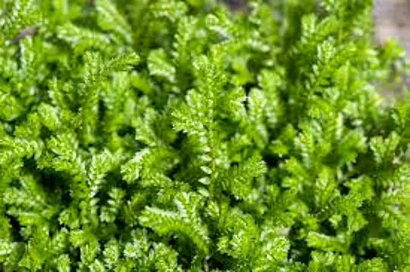
EXTERNAL FEATURES OF SELAGINELLA (SMALL CLUB MOSS)
- Many species are prostrate, creeping on the ground e.g. S. kraussiana, others are sub-erect e.g. S. trachyphylla or erect e.g. S. erythropus. A few species climb with the help of rhizophores e.g. S. alligans.
- The plant body is divided into root, stem and leaves.
- The primary root is short lived and all other roots are adventitious.
- On the basis of nature of stem and form of the leaves, the genus is sub-divided into two sub-genera-the homoeophyllum and the heterophyllum.
- In homoeophyllum species (S. selaginoides, S. rupestris, etc.) the stem is upright and all leaves are alike, while in heterophyllum species (majority of the species), the stem is prostrate and dorsiventral; and leaves are dimorphic (small and large).
- In homoeophyllum, all the leaves are alike, spirally arranged, small and simple.
- In heterophyllous species, they are dimorphic and are borne in pairs on dorsi ventral stem. The two leaves are markedly different in size (one is larger and other smaller).
- The smaller leaf of each pair is inserted on the dorsal side of the stem while the larger leaf is inserted on the ventral side.
- The successive pairs of leaves are so arranged, that large leaf always alternates with the large leaf, and small leaf with the small leaf.
- Each leaf is sessile, generally obovate with acute apex, and has a distinct midrib.
- At the base of each young leaf, on the adaxial face, their is small tongue-like out growth, the ligule.
- It is differentiated into basal sheath, glossopodium and the body of the ligule.
- Whereas the cells of the sheath are tubular in shape and are dead, those of the glossopodium are vertically elongated.
- The body of the ligule has parenchymatous cells with dense protoplasm.
- From the point where stem branches, a cylindrical leafless organ is seen growing downward. This is known as rhizophore.
- On reaching the ground, rhizophore terminates into roots (The morphological nature of the rhizophore is still open to question).
- Certain vertical branches from the stem are reproductive in nature and bear strobili.
Features of special interest
- Presence of rhizophore.
- Dimorphic leaves (in heterophyllous species).
- Presence of ligule.
ANATOMY OF ROOT OF SELAGINELLA (SMALL CLUB MOSS)
- The section is almost circular in outline.
- The tissues are differentiated into epidermis, cortex and stele.
- The epidermisis single layered and its cells are tangentially elongated. Few of these cells give rise to the root hairs.
- Cortexmay either be made of parenchymatous (thin walled) cells or the outer layer of cells may form a sclerenchymatous (thick walled) hypodermis.
- The stelelies in the centre. It is protostelic, monarch and exarch.
- Endodermisis one indistinct. layered and generally
- PericycIeis one to three layered.
- Xylemforms only one group. Protoxylem is situated towards the periphery.
- Phloemsurrounds the centrally located xylem.
ANATOMY OF RHIZOPHORE OF SELAGINELLA (SMALL CLUB MOSS)
- The outline of the section is almost circular.
- The section shows epidermis, hypodermis, cortex, endodermis and stele.
- The epidermisis cuticularised.
- Hypodermisthat follows is 2-3 celled thick.
- Cortexis few celled and parenchymatous. It occupies most of the part of section.
- Endodermisis present between the stele and the cortex. It is followed by a single layered parenchymatous peri cycle.
- The steleis a protostele. It shows monarch and exarch condition. In some species (e.g. s. atroviridis) the metaxylem is lunar shaped and many protoxylem groups are situated on the concave adaxial side.
ANATOMY OF STEM OF SELAGINELLA (SMALL CLUB MOSS)
- The outline of the section appears slightly wavy.
- The section shows epidermis, cortex and the stele.
- Epidermisis the outermost layer. It is cuticularised and lack stomata.
- The cortexconsists of parenchymatous cells, without any intercellular spaces. All the cells of the cortex are thin walled.
- Hypodermisoccurs close to epidermis. It develops from cells of outer cortex which become thick walled. In xerophytic, species (e.g. S. rupestris, S. lepidophyUa) hypodermis is more thickened.
- The steleis generally a protostele.
- Endodermisseparates vascular tissue from the cortical region, by radially elongated endodermal cells, called as trabeculae, with conspicuous intercellular spaces between two trabeculae. In spite of their great elongation, trabeculae still retain the transverse thickenings, the casparian strips, on their radial walls, characteristic of endodermal cells. Xerophytic species lack trabeculae (e.g. S. lepidophylla and S. rupestris).
- Pericycleis a single layer surrounding the xylem and phloem and follows endodermis.
- Stele :-The number of steles in a stem varies from 1-16 thus exhibiting a polystelic condition.
- Single stele, when present is generally diarch and exarch.
- In S. kraussiana, the commonest species, there are two steles, each with a single exarch mass of protoxylem.
- The protoxylemmasses of the two steles point in opposite directions.
- The phloemconsists of smaller cells with dense protoplasm and completely surrounds the central core of xylem, in each stele.
Features of special interest
- Presence of modified endodermis in the form of trabeculae.
- Presence of more than one stele i.e. polystelic condition.
ANATOMY OF LEAF OF SELAGINELLA (SMALL CLUB MOSS)
- The section shows a slightly bulged midrib in the centre and the wings.
- It shows definite upper and lower epidermis, usually undifferentiated mesophyll and a central vascular bundle.
- The epidermis is one layered. The stomata are generally present on the abaxial surface (lower) but may also be present on the adaxial surface (upper), or on both the surfaces.
- The mesophyll is usually not differentiated into palisade and spongy parenchyma. It shows many conspicuous intercellular spaces.
- The cells of mesophyll contain chloroplasts, each of which has several pyrenoid-like bodies.
- The vascular bundle is concentric with xylem surrounded by phloem and is bounded by a bundle sheath.
SPORE PRODUCING ORGANS
- The spore producing organs are sporangia, aggregated in strobili which are generally present at the apices.
- In some cases (as exemplified by S. patula) the axis may grow beyond the strobilus, terminating into a vegetative shoot or even in a second strobilus.
- s. of the strobilus shows a strobilar axis, around which sporophylls are spirally arranged. Each sporophyll is ligulate and similar to a foliage leaf.
- The sporangia are of two types, borne in the axils of the sporophylls, attached either strictly to the axils or to the axis just above.
- Selaginella is heterosporous, with megaspores (large) and microspores (small), borne in their respective sporangia, known as megasporangia and microsporangia.
- If a micro sporangium is borne in the axil of the sporophyll, it is known as a microsporophyll but if it is a megasporangium, the sporophyll is termed as a megasporophyll.
- Generally strobilus bears both types of sporangia but in S. gracilis, there are only one type of sporangia (either mega-or micro sporangia).
- When both kinds of sporangia occur in one and the same strobilus, their arrangement differs from species to species:
- In some species (e.g. S. oregana) there are only megasporangia on one side and only microsporangia on the other.
- In most of the species (e.g. S. kraussiana) there are only one or two megasporangia at the base and the rest are microsporangia.
- Both types of sporangia are stalked and have. two layered jackets. The outer layer of the jacket is chlorophyllous and has columnar cells, whereas the inner layer has tangentially elongated cells. It may form tapetum.
- The cells of the outer jacket are thickened, except at the apex.
- The two types of sporangia when ripe, differ -in their size, form, structure and colour.
- The megasporangium is much larger, four lobed, pale green or orange in colour and has only four megaspores.
- The microsporangium is smaller with uniform outline. It is dark brown or red in colour and has many spores.
- The megaspores are large in size and posses a triadiate ridge at its apex. It has thick sculptured exine and thin uniform intine.
- The microspores are pyramidal in shape, and have thick, ornamented exine and a thin, uniform intine. Both types of spores have a nucleus suspended in a rich cytoplasm.
IDENTIFICATION
- DIVISION – Pteridophyta
- True roots generally present (except in Psilopsida),
- True vascular strand present.
- Sub-division:- Lycopsida
- Laves microphyllous.
- Sporangia borne singly on adaxial face of the sporophyll or in its axil.
- Sporophylls borne in strobili.
- Order– Selaginellales
- Each foliage leaf with a ligule at the base on adaxial side.
- Sporophytes heterosporous
- Family – Selaginellaceae
- Stem herbaceous and dorsiventral or erect.
- Gametophytes extremely reduced.
- Genus – Selaginella
- Roots arise from rhizophore.
- Trabeculae present.
- Stele generally a protostele, sometimes siphonostele.
REFERENCES
- https://pixabay.com/photos/moss-selaginella-apoda-1526040/
- https://en.wikipedia.org/wiki/Selaginella
Leave a Reply