Contents
- 1 CLASSIFICATION OF PTERIDIUM (BRACKEN FERN)
- 2 EXTERNAL MORPHOLOGY OF PTERIDIUM (BRACKEN FERN)
- 3 ANATOMY OF ROOT OF PTERIDIUM (BRACKEN FERN)
- 4 ANATOMY OF RHIZOME OF PTERIDIUM (BRACKEN FERN)
- 5 ANATOMY OF RACHIS OF PTERIDIUM (BRACKEN FERN)
- 6 ANATOMY OF PINNULE OF PTERIDIUM (BRACKEN FERN)
- 7 STRUCTURE OF THE SPOROPHYLL OF PTERIDIUM (BRACKEN FERN)
- 8 STRUCTURE OF PROTHALLUS OF PTERIDIUM (BRACKEN FERN)
- 9 PROTHALLUS WITH YOUNG A SPOROPHYTE OF PTERIDIUM (BRACKEN FERN)
- 10 IDENTIFICATION OF ADIANTUM (MAIDEN HAIR FERN)
- 11 REFERENCES
CLASSIFICATION OF PTERIDIUM (BRACKEN FERN)
Kingdom :- Plantae
Division :- Pteridophyta
Sub-division :- Pteropsida
Class :- Leptosporangiatae
Order :- Filicales
Family :- Polypodiaceae
Genus :- Pteridium
Pteridium is cosmopolitan. It is widely distributed along the entire Himalayan tract. It grows particularly well at altitudes between 1,000 to 3,000 meters. P. aquilinum is found on forest floors, mountain slopes, open grasslands, etc.
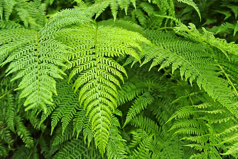
EXTERNAL MORPHOLOGY OF PTERIDIUM (BRACKEN FERN)
- The plant body is a sporophyte. It is differentiated into roots, rhizome and leaves.
- Stem is modified to rhizome. It is subterranean. The rhizome is long, slender and dichotomously branched. It is covered with brown and multicellular hairs called ramenta.
- The rhizome gives out adventitious roots on its underside. These are small and branched.
- The leaves are borne alternately on the upper side of the rhizome at the nodes.
- The young leaves are circinately coiled. The rachis is covered with ramenta.
- Each leaf is tripinnately compound. Each pinna is sessile. It has a distinct midrib that gives out lateral branches.
ANATOMY OF ROOT OF PTERIDIUM (BRACKEN FERN)
- The outline of the section is almost circular.
- It shows three regions-epiblema, cortex and the vascular cylinder.
- Epiblema is the outermost single layer of cells. The cells are thin walled and produce unicellular root hairs.
- Cortex occupies most part of the section It is differentiated into outer and inner regions.
- The outer region is parenchymatous while the inner few layers are sclerenchymatous.
- Endodermis follows the cortex. The radial walls of endodermal cells are characterised by casparian thickenings.
- Pericycle is situated inner endodermis. It is 1 or 2 layered and parenchymatous.
- Vascular cylinder shows radial, diarch and exarch conditions.
- The xylem consists of two central metaxylem tracheids with groups of small protoxylem elements on their both sides.
- Phloem is present on both the sides of xylem plate.
ANATOMY OF RHIZOME OF PTERIDIUM (BRACKEN FERN)
- The outline of the section appears almost like a biconvex lens.
- The tissues are differentiated into epidermis, hypodermis, ground tissue and the stele.
- Epidermis is the outermost single layer of cells. The cells are thickly cuticularised.
- Hypodermis lies below the epidermis. The cells are sclerenchymatous which often show pitted walls. It is generally interrupted on the lateral sides by parenchyma.
- Ground tissue follows the hypodermis. It is parenchymatous and is spread up to the center of the section. The cells are filled with starch grains.
- The structure of the stele varies with the age of the rhizome.
- In just formed rhizome, condition is protostelic.
- In a few weeks old plant with 2-3 leaves, the rhizome shows ectophloic siphonostele.
- In mature plant. the old part of rhizome shows a dictyostele.
- Dictyostele is made of meristeles arranged in two rings, separated by two sclerenchymatous bands.
- Meristele is surrounded by its own endodermis, which is followed by one or two layers of parenchymatous pericycle.
- The centre of the meristele is occupied by xylem which is completely surrounded by phloem on all sides.
ANATOMY OF RACHIS OF PTERIDIUM (BRACKEN FERN)
- The outline of the section appears horse-shoe shaped or hemispherical.
- The tissues of the section are differentiated into epidermis, hypodermis, ground tissue and the stele.
- Epidermiswhich is the outermost single layer of cells is thickly cuticularised.
- Hypodermisis present below the epidermis. It is 2 to 3 layered thick. The cells are sclerenchymatous.
- Ground tissue :-Following the hypodermis is a large region of parenchyma called ground tissue.
- Stele :-In the ground tissue is situated U-shaped or horse-shoe shaped stele.
- Endodermis and pericycle :-Stele is surrounded by a single layered endodermis followed by a few layered parenchymatous pericycle.
- Xylem :-The center of the stele is occupied by massive xylem. Metaxylem is present in the center with protoxylem located at
- Phloem :-The region between xylem and the pericycle is filled by phloem.
- The nature of the stele varies with the maturity of the rachis.
- In younger parts stele is U-shaped.
- Little above the base, it gets dissected into two large meristeles.
- In mature parts, many meristeles are present as a result of further dissection of the original stele.
ANATOMY OF PINNULE OF PTERIDIUM (BRACKEN FERN)
- The section shows the ‘midrib’ region and the wings.
- The midrib region consists of compact parenchyma in which a single concentric vascular bundle is situated. It shows centrally located xylem surrounded by phloem. A distinct parenchymatous bundle sheath surrounds the bundle.
- The upper and the lower epidermis are single layered. The stomata are present only on the lower surface.
- Mesophyll that lies between the two epidermal layers is differentiated into palisade and spongy parenchyma.
- The spongy tissue is situated close to the lower epidermis. The cells are loosely arranged and contain many chloroplasts. The intercellular spaces open into stomata.
STRUCTURE OF THE SPOROPHYLL OF PTERIDIUM (BRACKEN FERN)
- The leaf bearing sori is called sporophyll.
- The sporangia occur in groups called sori on the lower or abaxial side of pinnules. The sporangia form a continuous linear sorus along the margins. Such a confluent sorus is called coenosorus. The identity of sorus is thus lost and only one long sorus appears along the two lateral margins of the fertile pinnules.
- The sorus is protected by indusium. It is made of upper indusial flap formed by the incurved margins of the pinnule and the lower true indusial flap that is poorly developed.
- The sporangia in the sorus occur mixed. The development is leptosporangiate.
- Each mature sporangium is differentiated into a stalk and a capsule.
- The stalk of the capsule is made of three rows of cells. It is long and slender.
- The capsule is ovate or biconvex. The sporangial jacket is single layered thick. A ring . of thick walled cells forms The annulus. A few thin walled cells of the ring fonn the stomium. The capsule wall encloses 32 or 64 spores.
- All the spores being similar, the fern is homosporous. Spores are haploid and uninucleate. The wall is two layered. The outer thick layer is called exine and the inner thin layer is called intine.
STRUCTURE OF PROTHALLUS OF PTERIDIUM (BRACKEN FERN)
- The prothallus is a gametophyte formed as a result of spore germination.
- It is dark green, heart-shaped and single layered sheet of cells. The midrib region becomes a cushion of several cells. It remains attached to the substratum by rhizoids produced on the lower side in the central region.
- The antheridium is surrounded by the cells of the prothallus. Each antheridium consists of wall of three rings of cells. It encloses 30-40 multiflagellate antherozoids at maturity.
- Archegonia develop near the apical notch. Each is made of neck and ventre. The neck is 5-7 celled high with a single binucleate neck canal cell. The ventre has a small ventre canal cell and a large egg.
PROTHALLUS WITH YOUNG A SPOROPHYTE OF PTERIDIUM (BRACKEN FERN)
- Sporophyte is formed as a result of fertilization. The zygote grows into a sporophyte that still remains attached to the prothallus.
- Young sporophyte is differentiated into young leaves, primary and secondary roots.
- The leaves are petiolate and erect. These emerge through the apical notch. The leaves are simpler than the mature leaves. Sometimes these even show circinate vernation.
- Primary root grows on the lower side and gives out secondary roots.
- The sporophyte is dependent on the gametophyte till first leaf is formed. It absorbs its food through the foot of the young embryo.
IDENTIFICATION OF ADIANTUM (MAIDEN HAIR FERN)
- DIVISION – Pteridophyta
- True roots generally present (except in Psilopsida),
- True vascular strand present.
- Sub-division:- Pteropsida
- Vascular cylinder siphonostelic, with leaf gaps.
- Plants macrophyllous, leaves compound, with rachis.
- Leaves bear sporangia in sori.
- Gametophytes small, green and free living.
- Class:- Leptosporangiatae :-
- Sporangium with a jacket layer one cell in thickness.
- Definite number of spores.
- Order– Filicales
- Sori are mixed
- Family – Polypodiaceae
- Annulus of sporangium vertical,
- Each sporangium witb 32-64 spores.
- Genus – Pteridium
- Leaves tripinnately divided.
- Presence of coenosorus.
- Sorus enclosed between indusial flaps.
REFERENCES
- https://en.wikipedia.org/wiki/Bracken
- https://www.istockphoto.com/search/2/image?phrase=PTERIDIUM%20
Leave a Reply