Contents
- 1 CLASSIFICATION OF PINUS (PINE)
- 2 EXTERNAL FEATURES OF PINUS (PINE)
- 3 ANATOMY OF YOUNG ROOT OF PINUS (PINE)
- 4 ANATOMY OF OLD ROOT OF PINUS (PINE)
- 5 ANATOMY OF THE YOUNG LONG SHOOT PINUS (PINE)
- 6 ANATOMY OF THE OLD LONG SHOOT OF PINUS (PINE)
- 7 STUDY OF R.L.S. OF THE WOOD OF PINUS (PINE)
- 8 STUDY OF T.L.S. OF WOOD OF PINUS (PINE)
- 9 T.S. OF DWARF SHOOT AT THE BASE (BEFORE SECONDARY GROWTH) OF PINUS (PINE)
- 10 T.S. OF DWARF SHOOT AT BASE OF PINUS (PINE)
- 11 T.S. OF THE DWARF SHOOT AT UPPER END OF PINUS (PINE)
- 12 T.S. NEEDLE (LEAF) OF PINUS (PINE)
- 13 MALE CONE, MICROSPOROPHYLLS AND MICROSPORANGIA OF PINUS (PINE)
- 14 L.S. OF MALE CONE OF PINUS (PINE)
- 15 MORPHOLOGY OF THE FEMALE CONE OF PINUS (PINE)
- 16 L.S. OF FEMALE CONE PINUS (PINE)
- 17 L.S. OF OVULE OF PINUS (PINE)
- 18 SEED OF PINUS (PINE)
- 19 SEEDLING OF PINUS (PINE)
- 20 IDENTIFICATION OF PINUS
CLASSIFICATION OF PINUS (PINE)
Kingdom :- Plantae
Division :- Gymnosperm
Class :- Coniferopsida
Order :- Coniferales
Family :- Pinaceace
Genus :- Pinus
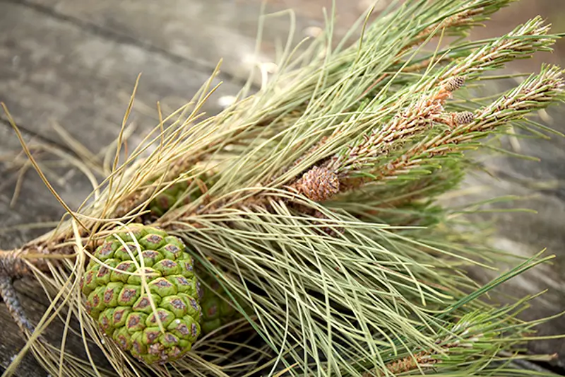
EXTERNAL FEATURES OF PINUS (PINE)
- It is a tall conical tree and, therefore, commonly grouped under conifers.
- The plant bodyis differentiated into root, stem and leaves.
- Underground root systemis formed by tap roots which disappear early and only lateral roots persist later on.
- The younger rootsare generally surrounded by fungal hyphae- the ectotrophic mycorrhizae.
- Aerial branch systemconsists of cylindrical, rough (being covered with scaly bark) and branched stem.
- The branchingis monopodial and the branches are arranged in whorls.
- Thebranches are dimorphic (of two types)- branches of unlimited growth or long shoots and branches of limited growth or dwarf shoots.
- Branches of unlimited growth or long shoots are present on the main trunk. These are produced at regular intervals.
- Branches of limited growth or dwarf shoots are borne on the main stem and on long shoots in the axils of scale leaves. Dwarf shoots also possess many scale leaves and bear group of foliage leaves at the apex.
- The leavesare also dimorphic (of two types)- scale leaves and foliage leaves.
- Scale leavesare brown, membranous and small. They are present on both the types of branches (i.e. long and dwarf shoots)
- Foliage leavesare green, acicular and needlelike. They are borne only by the dwarf shoots.
- A dwarf shootwith a group of needle-like foliage leaves is known as a foliar spur. The number of needles in a group varies from species to species. P. monophylla has a single leaf and spur is known as monofoilar, while in P. sylvestris, two leaves are present and spur is called as bifoiiar. In P. longifolia and P. gerardiana, they are three in number, the spur being called as trifoliar. Quadrifoilar spur occurs in P. quadrifolia and pentafoliar in P. excelsa.
- The shape of the needlevaries, according to their number in a spur. In P. sylvestris (with bifoliar spur), single needle is semi-circular in T.s. while in P. longifolia (with trifoliar spur), single needle is almost triangular in shape.
- Pinus is monoecious. Plant bears male and female reproductive parts in cones on the same plant.
- The male conesare borne on lateral branches of unlimited growth. They are produced in clusters and replace the dwarf shoots. Also, they are formed earlier in the season than the female cones.
- The female conesare borne terminally on branches of unlimited growth. They are produced singly and replace the long shoot. The female cone appears after every three years.
- Generally male and female cones are not formed on one and same branch.
ANATOMY OF YOUNG ROOT OF PINUS (PINE)
- The section is almost circular in outline.
- The tissues are differentiated into epiblema cortex and vascular tissues.
- Epiblemais outermost single layer. It gives out many thin and unicellular root hairs.
- Cortexis multilayered and parenchymatous.
- Endodermisseparates outer cortex and central vascular cylinder. It is single layered and cells are radially thickened.
- Pericyclefollows endodermis. It is multilayered
- Vascular bundlesare radial, exarch and diarch to hexarch.
- Protoxylemis generally Y-shaped and a resin canal is present in between the arms of Y.
- Pithis very small and lies between the groups of xylem.
ANATOMY OF OLD ROOT OF PINUS (PINE)
- The sectionshows cork, cortex, primary and secondary vascular tissues and a small pith.
- Corkforms the outermost several layers. (developed from pericycle and hence primary cortex is completely peeled off).
- Stone cells occur in many groups scattered just below the zone of cork.
- Secondary cortexfollows cork. It is parenchymatous and a few layered deep.
- Many resin canalsare found in the secondary cortex.
- Primary phloemoccurs in two patches. The tissues are mostly crushed and obliterated.
- Secondary phloemthat follows is a few layered deep ring. It consists of sieve tubes, sieve plates, phloem parenchyma and albuminous cells.
- Secondary phloem and secondary xylem are separated by a cambium.
- Secondary xylemis composed of tracheids arranged in regular rows. It is traversed by uniseriate medullary rays.
- Pithis small and parenchymatous. Two groups of primary xylem are situated on opposite radii.
- Each primary xylemgroup is Y shaped. The divided arm faces the outer side (away from the pith).
- Resin canal :-The characteristic of the pine root is the presence of large resin canal between the divided arm of Y, close to each primary protoxylem group.
ANATOMY OF THE YOUNG LONG SHOOT PINUS (PINE)
- Outlineis wavy due to the presence of scaly leaves.
- The stemis differentiated into epidermis, cortex and stele.
- Epidermisis the outermost single layer. It is thickly cuticularized.
- Cortexis multilayered and lies below the epidermis. The outer few layers forming hypodermis are sclerenchymatous. Inner layers are thin walled and parenchymatous in which large number of resin canals and leaf traces are distributed irregularly.
- Resin canal :-The cavity of resin canal is bounded by a glandular, resin secreting epithelial layer. Outer to this layer are one or two layers of sclerotic cells.
- In Pinus, resin canalsare present in the cortex and secondary wood of both stem and root and on margins of the primary xylem in the root.
- The steleis ectophloic siphonostele.
- Endodermisis present but is undistinguishable and so also a few layered peri cycle located inner to it.
- Vascular cylinderis composed of 5-8 vascular bundles, separated by medullary rays. Vascular bundles are arranged in a ring.
- Each vascular bundleis conjoint, collateral endarch and open.
- Xylemis composed of tracheids and xylem parenchyma only, vessels are absent.
- The phloemis made up of sieve tubes, sieve plates and phloem parenchyma. Albuminous cells are also present.
- Pith lies in the centre and is parenchymatous. It is connected with the cortex but narrow medullary rays separate the vascular bundles.
ANATOMY OF THE OLD LONG SHOOT OF PINUS (PINE)
- The sectionshows cork, cortex, primary and secondary vascular tissues and pith.
- Cork :-The outmost region is formed by the successive layers of cork. It consists of thick and suberized cells.
- Cork cambiumfollows cork. It is made of a few layers of regularly arranged cells.
- Secondary cortexpresent below is parenchymatous.
- Primary cortexis parenchymatous and many layered. The resin canals occur irregularly distributed in this region.
- Primary phloemthat lies inner to primary cortex occurs as small patches of crushed tissues.
- Secondary phloemoccurs as a well distinguished ring.
- Phloemis composd of sieve tubes and phloem parenchyma.
- Cambiumseparates the secondary phloem on its outer side and secondary xylem on its inner side.
- Secondary xylemshows distinct and sharp annual rings. Thin walled and large xylem elements form a ring of spring wood. Thick walled and small xylem elements form a ring of autumn wood. The wood is pyconoxylic (compact).
- Rings of secondary xylem– autumn and spring wood alternate one another and together form annual ring.
- Secondary xylem(wood) is composed of tracheids and ‘xylem parenchyma. Vessels are completely absent. Hence it is called nonporous wood.
- Medullary raystraverse xylem and phloem. Primary medullary rays run from primary xylem to secondary phloem.
- Primary xylemgroups are endarch and lie just near the pith.
- Resin canalsare scattered in the primary and secondary xylem as in the cortex.
- Pithis small, parenchymatous and many cells are filled with tannin.
STUDY OF R.L.S. OF THE WOOD OF PINUS (PINE)
- It shows presence of secondary xylem, ray tracheids and medullary rays.
- Xylemis composed of tracheids with bordered pits on their radial walls. The bordered pits in this section are seen in surface view
- Bordered pitsare circular areas surrounded by special cellulose thickenings called crassulae or Bars of Sanio. If pits are close to one another, the bars fuse to form Rims of Sanio.
- Medullary raysrun horizontally. In radial longitudinal plane they are cut length-wise and their length and height can be noticed. They are uniseriate.
- Each medullary rayis made up of ray cells, ray tracheids and parenchyma.
- Ray tracheidsare present on both the sides of the medullary ray cells, only in the region of xylem. These cells are thick, narrow and long. They show bordered pits.
- Ray parenchymaoccurs between the tracheids. These cells are thin, broad, small and living.
- Medullary ray, in the region of phloem replaces ray tracheids with albuminous cells. They are small and contents are dense. (Ray parenchyma associated with these cells is filled with large amount of starch).
STUDY OF T.L.S. OF WOOD OF PINUS (PINE)
- Tracheids and medullary rays are cut transversely in this plane.
- The bordered pitsare cut to show ovearching borders, forming a dome-like structure. It encloses in the centre a small disc, called torus.
- Medullary raysare uniseriate. Since they are cut transversely, their height and breadth can thus be determined.
- Each medullary ray appears to be a short row of more or less rounded cells, three or four cells high.
- Composition of medullary ray reveals centrally placed, thin-walled and living cells-the albuminous cells (in the phloem region) and the ray cells (in the xylem region).
- These are surrounded on the lower and upper sides by thick walled and dead cells known as ray tracheids.
T.S. OF DWARF SHOOT AT THE BASE (BEFORE SECONDARY GROWTH) OF PINUS (PINE)
- The section almost resembles with that of the main stem.
- The outlineis wavy, due to ensheathing scaly leaves.
- The tissuesare differentiated into epidermis. cortex and stele.
- Epidermisis made of single layer of thick walled cells.
- Cortexfollows epidermis. Outer few layers, close to epidermis are thick walled, while the inner layers are thin walled and parenchymatous.
- Resin canals are present in the cortex. These are about six in number. Tannin cells are also irregularly scattered in this region. 7. Stele is an ectophloic siphonostele.
- Endodermisis single layered and is followed by pericycle. Both the layers are indistinguishable.
- Vascular bundlesvary in number. They are generally six. Each vascular bundle is conjoint, collateral, endarch and open.
- Pithis small. The cells are thick walled.
- Medullary raysconnect the pith and cortex and separate vascular bundles from one another.
T.S. OF DWARF SHOOT AT BASE OF PINUS (PINE)
- Dwarf shoot also shows a little amount of secondary growth.
- The sectionshows scale leaves, single-layered thick walled epidermis, few layers of cork cells and tannin-filled cells.
- Primary phloemis crushed and form patches.
- Secondary phloemunderlies it and forms a complete ring.
- Secondary xylemis small and is separated by a thin ring of cambium from the phloem region. Medullary rays traverse the secondary xylem.
- Endarch protoxylemgroup lies just near the pith. It is small and consists of thick-walled cells. Few cells are tannin-filled.
T.S. OF THE DWARF SHOOT AT UPPER END OF PINUS (PINE)
- The structure is essentially similar to one found at its base.
- The tissues of the dwarf shoot, towards the upper part, gradually become separated into equal parts, corresponding to the number of leaves in a spur (e.g. Pinus gerardiana, with trifoliar spur shows division of the dwarf shoot into three equal parts while in P. quadrifolia, with quadrifoliar spur, gets separated into four equal parts).
- Each part shows distinct epidermis with stomat~ present all over.
- Parenchymatous cortex fills most part of the section. Resin canals are located in the comers.
- In the center two conjoint, collateral and endarch vascular bundles are present. These are surrounded by distinct endodermis and pericycle.
T.S. NEEDLE (LEAF) OF PINUS (PINE)
- The outlineof the section varies according to the species. (Triangular if spur is trifoliar, semicircular if spur is bifoliar)
- The needleis differentiated into epidermis, mesophYll and stele.
- Epidermisis single with tangentially elongated and thickly cuticularized cells.
- Stomataare sunken. These are present on all the faces of epidermis. The needle is thus said to the amphistomatic.
- Epidermisis followed by hypodermis. It is few layered thick at the corners and 1-2 layered in other parts. Sub-stomatal chambers occur in this region. Cells are sclerenchymatous and fibrous.
- Mesophylllies below the hypodermis. It is made up of polygonal parenchymatous cells, densely filled with the chloroplasts. Numerous plate-like or peg-like infoldings project into the cell lumen (cavity) from the wall of the mesophyll cells.
- Resin canalsgenerally occur in the sclerotic hypodermis but also occur in the mesophyll tissue.
- Endodermisis conspicuous. Cells are barrel shaped and tangentially thickened. It is followed by a many layered, parenchymatous pericycle.
- . Generally two vascular bundles remain surrounded by this tissue. (In P. strobus there is only one vascular bundule).
- Thevascular bundles are separated from one another by a T-shaped thick walled transfusion tissue.
- Each vascular bundleis conjoint, collateral and open. Protoxylem faces adaxial side. Phloem is located on the abaxial side.
- Xylem and phloemgroups are separated from one another by cambium at the base of the needle and by parenchymatous cells in the upper region.
- Secondary growthis very little during which the medullary rays run between xylem and phloem.
Features of special interest
It shows the following xerophytic characters
- Narrow acicular form of the leaf.
- Presence of thick cuticle.
- Aphistomatic nature.
- Sunken stomata.
- Thick and sclerenchymatous hypodermis.
- Infolded peg-like structures in mesophyll.
- Presence of transfusion tissue.
- Simple vascular system.
MALE CONE, MICROSPOROPHYLLS AND MICROSPORANGIA OF PINUS (PINE)
- Male cones replace the dwarf shoots. Each male cone arises in the axile of a scale leaf. The main shoot, on which these are produced, continues to grow further.
- Male cones are grouped in clusters on the shoots of the same year only.
- Each male cone has single, centrally located cone axis around which many scaly microsporophylls are spirally arranged.
- Each microsporophyll has an expanded triangular central part and stalk-like base. Terminal part projects into a tip.
- Few lowermost sporophylls are sterile, and do not bear any male reproductive structures.
- On the abaxial side, each microsporophyll bears two ovoid micro sporangia or pollen sacs on its lateral sides.
- Each micro sporangium has its own wall which encloses many microspores
- The young microspore is globular or spherical in shape and is uninucleate.
- A mature microspore or pollen grain shows two wall layers- exine and intine, 2 prothallial cells and antheridial cell.
- Pollen grain has a thick expanded exine in the form of wings on the sides, followed by a smooth intine.
L.S. OF MALE CONE OF PINUS (PINE)
- It shows a cone axis bearing microsporophylls.
- The cone axis is centrally located.
- Microsporophylls are spirally arranged. These are scaly, triangular and expanded.
- It is attached to the cone axis by a stalk-like base.
- The outer expanded part is sterile and is known as apophysis.
- Microsporangia are present on the lower or abaxial surface.
- Each micro sporangium has a wall that encloses a cavity.
- The wall consists of epidermis. wall layers and tapetum.
- The cavity shows numerous microspores in various stages of development.
MORPHOLOGY OF THE FEMALE CONE OF PINUS (PINE)
- Female cones are larger than the male cones. They are borne at the apices of the young elongated shoots, replacing the shoot of unlimited growth (long shoots).
- Single shoot may bear one to four female cones which are reddish-green in colour and mature in three years.
- In the first year, cones are compact and sporophylls are closely arranged.
- The second year cones are large in size and woody in nature but sporophylls are still compactly arranged.
- In the third year, cone becomes loose. Sporophylls separate from one another due to elongation of the cone axis.
- Each female cone consists of many sporophylls, arranged spirally around the cone axis.
L.S. OF FEMALE CONE PINUS (PINE)
- Female cone is made of centrally located cone axis and spirally arranged sporophylls.
- Each sporophyll consists of two kinds of paired scales :
- bract scale or cone scale and
- ovuliferous scale or seminiferous scale.
- Many small and thin bract scales are arranged spirally around the cone axis. They are directly borne on the cone axis. Each of these is present on the abaxial (lower) side of the ovuliferous scale.
- On the adaxial (upper) side of the bract scale, a thick, large, woody and triangular ovuliferous scale is present.
- The ovuliferous scales in the middle part of the cone are the largest and get gradually smaller towards its base and apex.
- Ovuliferous scale and bract scale are fused for a little distance near the cone axis while free at a distance away from it.
- Ovuliferous scale is shortly stalked and rest of the part is expanded.
- At the base “of this expanded, triangular part, two naked and sessile ovules are present. These are situated on the adaxial, (upper) surface of the ovuliferous scale, at its base, with their micropyles directed towards cone axis.
- The terminal part of the ovuliferous scale is broad and sterile and is known as apophysis.
L.S. OF OVULE OF PINUS (PINE)
- Ovule is elongated in shape.
- It is unitegmic and the integument is three layered. The outermost layer is thin. The middle layer is stony and prominent. The innermost layer is fleshy and well developed.
- Nucellus is fused with inner layer of the integument, except at its tip where it forms an elongated and slender micropyle, directed towards the cone axis.
- In the nucellar region lies a small cavity just opposite the micropyle. It is known as pollen chamber.
- Female gametophyte (endosperm) is differentiated from nucellus. About 2-5 archegonia are situated in this region at the micropylar end near the base of the archegonial chamber
SEED OF PINUS (PINE)
- Fertilized ovules get transformed into seeds which are situated on the adaxial side of the ovuliferous scale at its base near the cone axis.
- Seeds are small, elongated and winged. The wing is a thin layer of tissue which splits off from the adaxial face of the ovuliferous scale. (Seed can be best studied by cutting longitudinal section of the seed of P. gerardiana; vern. chilgoza).
- The seed is covered with red and brown testa.
- Inner fleshy layer of the integument still persists. It is membranous, thin and papery, termed as tegmen.
- The nucellus is present as a thin layer and forms a nucellar cap at the micropylar end.
- The larger part of the seed consists of oily endosperm.
- The suspensor is long and becomes coiled. Embryo is differentiated into radicle, plumule and cotyledons (3-8 in number).
- In between the radicle and plumule, is present a well developed hypocotyl.
SEEDLING OF PINUS (PINE)
- Seedling shows three parts (i) roots (ii) hypocotyl and (iii) leaves.
- The roots are well branched and arise from the radicle.
- Hypocotyl gives rise to unbranched, slender and thin primary shoot.
- Leaves are green and needle-like which are borne in whorls on the primary shoot. These are cotyledonary leaves.
- The primary leaves or first spur shoots arise in the axils of some of these juvenile (cotyledonary) leaves and are borne in spiral series on the primary shoot.
IDENTIFICATION OF PINUS
- DIVISION – Gymnosperms
- (1) Absence of vessels.
- Ovules naked.
- Seeds attached with woody acales.
- Scales generally form a cone.
- Class:- Coniferopsida
- Leaves needle shaped.
- Wood pycnoxylic (compact).
- Presence of resin canals.
- Compact male and female cones.
- Non-flagellate male gametes.
- Seeds bilaterally symmetrical.
- Order– Coniferales
- Family – Pinaceace
- Resinous wood.
- Plants monoecious.
- Sporophylls spirally arranged.
- Microsporophylls with two microsporangia.
- Pollen grains winged
- Female cone woody
- Polyembryony present
- Seed dry and winged.
- Genus – Pinus
- Plants sporophytic and monoecious. Male and female reproductive organs in cones
- Branches dimorphic
- Long shoots with secondary xylem, annual rings are formed, wood pycnoxylic and resinous
- Dwarf shoots with a little secondary growth
- Leaves are of two types
- Scale leaves brown and membranous
- Foliage leaves are acicular, xerophytic, mesophyll cells with peg-like ingrowths, 2 resin canals and T-shaped transfusion tissue
- Male cones borne laterally, in clusters, microsporophyll bears two microsporangia on abaxial side
- Pollen grains winged
- Female cones borne single and terminal
- Bract scales and ovuliferous scales spirally arranged
- Two naked ovules on the adaxial side of the ovuliferous scale
- Seeds dry and winged.
Leave a Reply