Contents
- 1 CLASSIFICATION OF NEPHROLEPIS (SWORD FERN)
- 2 EXTERNAL MORPHOLOGY OF NEPHROLEPIS (SWORD FERN)
- 3 ANATOMY OF ROOT OF NEPHROLEPIS (SWORD FERN)
- 4 ANATOMY OF RHIZOME OF NEPHROLEPIS (SWORD FERN)
- 5 ANATOMY OF RACHIS OF NEPHROLEPIS (SWORD FERN)
- 6 STRUCTURE OF THE SPOROPHYLL OF NEPHROLEPIS (SWORD FERN)
- 7 IDENTIFICATION OF ADIANTUM (MAIDEN HAIR FERN)
- 8 REFERENCES
CLASSIFICATION OF NEPHROLEPIS (SWORD FERN)
Kingdom :- Plantae
Division :- Pteridophyta
Sub-division :- Pteropsida
Class :- Leptosporangiatae
Order :- Filicales
Family :- Polypodiaceae
Genus :- Nephrolepis
It is commonly found in tropics, but a few species like Nephrolepis acuta, N. tube rosa, etc. are also grown as ornamentals.
EXTERNAL MORPHOLOGY OF NEPHROLEPIS (SWORD FERN)
- The plant body IS a sporophyte. It is differentiated into roots, rhizome and leaves.
- The rhizome gives out adventitious roots from its underside. These adventitious roots are small and branched.
- The stem IS modified to rhizome. It is subterranean, short and erect. The rhizome produces elongated slender stolons. Peltate scales cover the rhizome.
- In N. tuberosa, rhizome bears tubers. These are reservoirs of carbohydrates and water.
- The leaves are long, narrow, sub-coriaceous and unipinnate.
- The pinnae are sessile or shortly petioled. They have a usually rounded or cordate base. Each pinna has articulation with a pouch-like structure at the base.
- The veins are prominent and the veinlets are branched with open ends. The tips of veinlets are gland dotted and they extend up to the margins.
ANATOMY OF ROOT OF NEPHROLEPIS (SWORD FERN)
- The outline of the section is almost circular.
- The tissues are differentiated into epiblema, cortex and the vascular cylinder.
- Epiblema is the outermost single layer of cells. The cells are unicellular and thin walled. A few root hairs are produced by this layer.
- Cortex forms the major part of the section. It shows outer parenchymatous region and the inner small sc1erenchymatous region.
- Endodermis follows the cortex and separates it from the vascular tissues. The cells of endodermis show casparian strips.
- Endodermis is followed by 1 or 2 layered parenchymatous pericyc1e.
- Vascular cylinder is represented by a radial, diarch and exarch vascular bundle.
ANATOMY OF RHIZOME OF NEPHROLEPIS (SWORD FERN)
- The outline of the section is almost biconvex.
- The section can be divided into epidermis, hypodermis, ground tissue and the stele.
- Epidermis is the outermost single layer of thickly cuticularised cells.
- Hypodermis that follows epidermis is made of a few sc1erenchymatous layers.
- Ground tissue. Rest of the tissue is called ground tissue. It is parenchymatous with numerous starch grains in the cells.
- The structure of the stele varies with the age of the rhizome.
- In youngest part of rhizome, it is protostele.
- In a few weeks old plant with a few leaves, the rhizome shows ectophloic siphonostele.
- The old part of rhizome shows a dictyostele.
- Dictyostele is made of two rings of meristeles, separated by two sc1erenchymatous bands.
- Meristele has its own endodermis and pericyc1e. The centre is occupied with xylem which is completely surrounded by phloem on all its sides .
ANATOMY OF RACHIS OF NEPHROLEPIS (SWORD FERN)
- The section appears horse-shoe shaped.
- It shows epidermis, hypodermis, ground tissue and the stele.
- Epidermis is made of single layer of thickly cuticularised cells.
- Hypodermis lies below the epidermis. The cells are sclerenchymatous.
- Ground tissue. The rest of the parenchymatous region extending throughout the section is called ground tissue.
- In the ground tissue is situated V-shaped or horse-shoe shaped stele.
- The stele is surrounded by a single layered endodermis followed by a few layered pericycle.
- Xylem and phloem. Centrally located xylem is surrounded by phloem on all sides.
- The structure of the stele differs at various levels of rachis-
- In younger parts, there is a single V-shaped stele.
- Little above the base, the V-breaks at the bottom, thereby producing two steles.
- In mature parts, dissection of the stele results in many meristeles.
STRUCTURE OF THE SPOROPHYLL OF NEPHROLEPIS (SWORD FERN)
- The leaf bearing sori is called sporophyll.
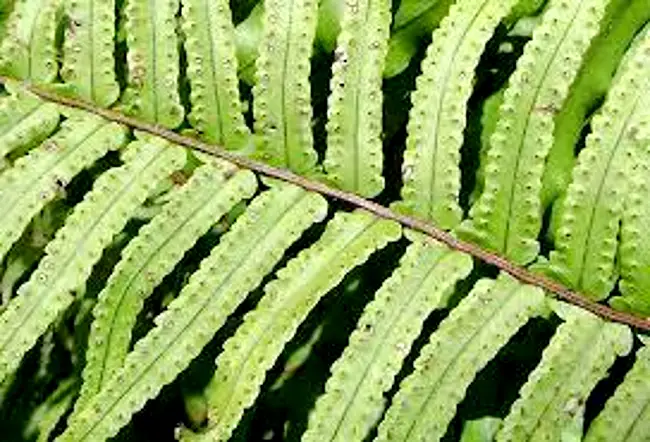
- The sporangia are present on the lower side of mature pinnae. These occur in groups called sori.
- Sori are superficial and form definite rows, one on either side of the vein.
- The sorus appears semi-rounded, and arises at the tip of the veinlet.
- The sori are indusiate. The indusium is reniform (kidney-shaped), roundish or sub-orbicular.
- Each sporangium has a stalk and a capsule.
- The stalk is long, slender and multicellular.
- The wall of sporangial capsule is one celled thick. A ring of thick walled cells called annulus is present. A few thin walled cells forming stomium are situated in the ring.
- The capsule wall encloses 32 or 64 spores. All the spores are similar, the fern being homosporous.
IDENTIFICATION OF ADIANTUM (MAIDEN HAIR FERN)
- DIVISION – Pteridophyta
- True roots generally present (except in Psilopsida),
- True vascular strand present.
- Sub-division:- Pteropsida
- Vascular cylinder siphonostelic, with leaf gaps.
- Plants macrophyllous, leaves compound, with rachis.
- Leaves bear sporangia in sori.
- Gametophytes small, green and free living.
- Class:- Leptosporangiatae :-
- Sporangium with a jacket layer one cell in thickness.
- Definite number of spores.
- Order– Filicales
- Sori are mixed
- Family – Polypodiaceae
- Annulus of sporangium vertical,
- Each sporangium witb 32-64 spores.
- Genus – Nephrolepis
- Leaves unipinnate with articulate or pouch like base.
- Sori distinct and enclosed by individual indusium
- Indusium true.
REFERENCES
- https://en.wikipedia.org/wiki/Nephrolepis_exaltata
- https://www.istockphoto.com/search/2/image?phrase=sword+fern


Leave a Reply