Contents
- 1 CLASSIFICATION OF MARSILEA
- 2 EXTERNAL MORPHOLOGY OF MARSILEA
- 3 ANATOMY OF ROOT OF MARSILEA
- 4 ANATOMY OF RHIZOME OF MARSILEA
- 5 ANATOMY OF PETIOLE OF MARSILEA
- 6 ANATOMY OF LEAFLET OF MARSILEA
- 7 EXTERNAL FEATURES OF SPOROCARP IN MARSILEA
- 8 T.S. OF SPOROCARP OF MARSILEA
- 9 V.L.S. OF THE SPOROCARP
- 10 H.L.S. OF THE SPOROCARP OF MARSILEA
- 11 DISPERSAL OF SPORES
- 12 IDENTIFICATION OF ADIANTUM (MAIDEN HAIR FERN)
- 13 REFERENCES
CLASSIFICATION OF MARSILEA
Kingdom :- Plantae
Division :- Pteridophyta
Sub-division :- Pteropsida
Class :- Leptosporangiatae
Order :- Marsileales
Family :- Marsileaceae
Genus :- Marsilea
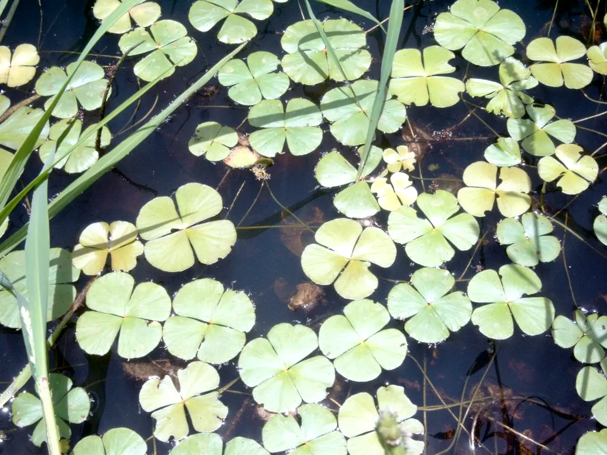
The two Indian species, Marsilea minuta and M. quadrifolia, are commonly found growing either in shallow water or on moist banks of ponds and ditches. They grow either completely submerged or partially or entirely out of water in damp and wet places.
EXTERNAL MORPHOLOGY OF MARSILEA
- The plant body is differentiated into a rhizome, roots and leaves.
- The rhizomeis slender, creeping branched. It may either grow in water or attached by roots in the damp soil.
- It bears nodes and internodes. The leaves and roots occur in acropetal succession (youngest towards the apex of rhizome) on the nodes. The adventitious roots grow downwards and the leaves grow upwards. Young leaves are circinately coiled, a characteristic of most ferns.
- The leavespresent at the nodes occur in two rows, (two ranked) one on either side of the mid-line of the rhizome.
- Each leaf consists of a long petiole, bearing at its top generally four leaflets or pinnae, apparently arising from one common point. In M. quadrifolia, a common Indian species, six leaflets are found. (Puri and Garg, 1953, call the leaflets as pinnules).
- The division of lamina into four pinnae is the result of three dichotomies, close to each other. Therefore, out of the four leaflets, two form a distal pair while the lower two are alternate. The leaflets, thus give a false impression of arising from one common point.
- Each leafletis obovate. The venation is dichotomous with several cross connections. The free veinlets at the apex of the leaflet are tied up with marginal loops.
- Leaflets fold up in the night or early morning, thus showing sleeping movements.
- The plant when grows in water, has long, flexible petioles and the leaflets float on the surface of the water but when it grows on mud or damp soil, the petioles become short and rigid. (It is interesting to note that, when in a pond in which Marsilea is growing, water level rises, the petioles are also seen to increase in length. Contrary to it when level goes down, the petioles are found to coil, as such in both the conditions, the leaflets float on water surface).
- The spore bearing structures known as sporocarps are commonly borne laterally near the base, on the petiole, but sometimes higher up. The two common Indian species, M. minuta and M. quadrifolia show variation in the number of sporocarps from one to four.
ANATOMY OF ROOT OF MARSILEA
- The outlineof the section appears almost circular.
- The epidermisis single layered with tangentially elongated cells.
- The cortexis differentiated into an outer and an inner cortex.
- The outer cortexhas many air chambers separated by radial septa.
- The inner cortexhas either all the parenchymatous cells or some of the cells towards the inner side may become thick walled and sclerenchymatous.
- Endodermisis single layered. It is followed by one layered pericycle. These surround vascular bundle.
- Xylemis diarch and exarch xylem. It is situated in the centre. The protoxylem elements are situated opposite one another.
- The phloemhas smaller cells and forms two bands, one on either side of the xylem mass. Features of special interest The root shows aerenchyma III the outer cortex (hydrophytic character).
ANATOMY OF RHIZOME OF MARSILEA
- The outlineof the section appears almost circular.
- The section shows three regions-epidermis, cortex and stele.
- The epidermisis single layered without stomata. The epidermis of aquatic plants lacks cuticle but that of terrestrial individuals has a distinct cuticle.
- The cortexis differentiated into three regions-the outer, the middle and the inner.
- The outer cortexhas well-developed air
- The middle cortexis thick walled, made up of sclerenchymatous cells and is only a few celled thick.
- The inner cortexis composed of thin-walled parenchymatous cells containing starch.
- The steleis an amphiphloic solenostele.
- Steleshows a central xylem ring. On its outer side is outer phloem ring. On the inner side i.e. towards the pith are present inner phloem ring, inner pericycle and inner endodermis. phloem
- Protoxylemgroups mayor may not be distinct. They are generally exarch, but in some cases mesarch too.
- Pithlies in the center. In aquatic plants it is parenchymatous and in terrestrial plants it is sclerotic.
Features of special interest
- It shows hydrophytic character viz. presence of aerenchyma in the cortex, as well as some xerophytic characters viz.
- thick walled middle cortex and
- sclerotic pith
- Presence of amphiphloic solenostele.
ANATOMY OF PETIOLE OF MARSILEA
- The outlineof the section is circular.
- Epidermisis the outermost layer with rectangular cells.
- Hypodermisis sometimes present below the epidermis. It is one or two layered.
- The cortexis differentiated into an outer and an inner zone.
- The outer cortexhas many air chambers, separated by narrow radially arranged parenchymatous cells (aerenchyma).
- The inner cortexhas parenchymatous cells containing starch. A few cells contain tannins also.
- The steleis a protostele.
- Endodermisis single layered. It is followed by a single layer of pericycle.
- The xylemis ‘V shaped’ with exarch protoxylem. The two arms of ‘V’ are slightly curved and separate. Each arm has generally one or two large tracheids in the middle and smaller tracheids towards both the ends. The open end of ‘V’ always points towards the adaxial side of the petiole (towards the axis)
- Phloem surrounds the xylem.
Features of special interest
- Shows hydrophytic character viz. presence of aerenchyma in the outer cortex
- Presence of V-shaped xylem.
ANATOMY OF LEAFLET OF MARSILEA
- The section shows an upper and lower epidermis, mesophyll and a vascular bundle.
- The stomataare found on both upper and lower epidermis if the plant is terrestrial but they are found only on upper epidermis if leaves float on water surface.
- Mesophyllis differentiated into palisade and spongy parenchyma.
- Palisadeis arranged in one layer near the upper epidermis. Spongy parenchyma is located near the lower epidermis. It is loosely arranged to form large air spaces and is called aerenchyma.
- There are many vascular bundles. Each bundle is concentric with centrally located xylem surrounded by phloem.
- The distinct endodermis is present just outside the vascular bundle
EXTERNAL FEATURES OF SPOROCARP IN MARSILEA
- The spore-bearing organs are the sporocarps which are borne laterally on the adaxial side of the petiole. Their number and positions vary from species to species.
- Sporocarp is stalked, bean-shaped or ovoid structure.
- The place of attachment of the body of the sporocarp to the peduncle (stalk) is known as raphe.
- Beyond the raphe, there are two projections known as teeth or tuberceles, one tooth being lower than the other.
- The lower tooth is usually stouter and more prominent while the upper tooth, which lies a short distance above is usually more slender and delicate.
- The side on which the raphe is present is the basal side and the side opposite to it is the apical side. The side on which the tubercles are present is the dorsal side and the side opposite to it is ventral side
T.S. OF SPOROCARP OF MARSILEA
- The section shows wall of the sporocarp which encloses sori.
- The wall is made of outer epidermis followed by hypodermis.
- Epidermis consists of thick walled cells. Numerous stomata are present in the epidermis.
- Hypodermis consists of two layers of radially elongated cells. The cells of the inner layer are double in length as compared to the cells of the outer layer.
- All the cells of both the layers have their nuclei arranged in one row.
- Receptacles are cut longitudinally. Only two sori are seen, each of which is covered by its own 2 layered indusium. The receptacle of the sorus bears microsporangia at the corners and megasporangia all along the receptacular ridge.
- On the upper and lower sides of the receptacles, two masses of gelatinous ring, cut transversely, are present. The upper one is bigger in size than the lower.
- The dorsal bundle, lateral bundles, placental branches and placental bundles are seen
V.L.S. OF THE SPOROCARP
- The section shows wall of the sporocarp enclosing sori embedded in a gelatinous wall.
- The outermost is the sporocarp wall. It is made of an epidermis with stomata and two layered hypodermis.
- Below the sporocarp wall is a gelatinous ring which surrounds sori. It is relatively more prominent on the dorsal side than on the ventral.
- The sori are cut longitudinally and appear in a row.
- Each sorus is surrounded by its own indusium.
- If the section passes through the centre, then megasporangia are seen in all the sori. Since the megasporangia are present at the apex of the receptacle, no receptacle is seen.
- If the section is not perfectly median, then microsporangia are seen attached on either sides of the receptacle in each sorus.
- In this section the stalk bundle and cut lateral bundles are seen.
H.L.S. OF THE SPOROCARP OF MARSILEA
- The section shows transversely cut stalk, wall of the sporocarp and two rows of sori.
- Transversely cut stalk appears on one side. It shows the stalk bundle.
- The wall of the sporocarp is made of epidermis with stomata and two layered hypodermis.
- Gelatinous ring shows two patches, heavier on the dorsal side than on the ventral.
- There are two rows of sori, one row alternating with the other.
- Each sorus is covered by its indusium.
- A sorus consists of a receptacle. Megasporangium is present at the apex of receptace while microsporangia are present on the sides.
- The lateral bundles are cut transversely and each is seen to supply its own receptacle by a receptacular or placental branch.
- Thus, in this section dorsal bundle, many lateral bundles and receptacular branches (placental branches) are seen.
DISPERSAL OF SPORES
- The sporocarp is hard and resistant to unfavourable conditions.
- It opens through its ventral margins.
- It imbibes water and the gelatinous ring inside swells up.
- This ring ultimately comes out of the sporocarp wall.
- Gelatinuous ring bears two rows of sporangia, one on each side, alternating with one another.
IDENTIFICATION OF ADIANTUM (MAIDEN HAIR FERN)
- DIVISION – Pteridophyta
- True roots generally present (except in Psilopsida),
- True vascular strand present.
- Sub-division:- Pteropsida
- Vascular cylinder siphonostelic, with leaf gaps.
- Plants macrophyllous, leaves compound, with rachis.
- Leaves bear sporangia in sori.
- Gametophytes small, green and free living.
- Class:- Leptosporangiatae
- Sporangium with a jacket layer one cell in thickness.
- Definite number of spores.
- Order– Marsileales
- Members heterosporous,
- Sporangia formed within sporocarps.
- Family – Marsileaceae
- Members aquatic.
- Sorus gradate
type and each produces both types of sporangia - Leaf circinately coiled in bud condition.
- Genus – Marsilea
- Leaflet with dichotomous venation and cross connections,
- Presence of aerenchyma in vegetative organs of the sporophyte.
- Presence of amphipbloic solenostele in the rhizome.
- Presence of V-shaped xylem in petiole.
REFERENCES
- https://en.wikipedia.org/wiki/Marsilea
- https://depositphotos.com/stock-photos/marsilea.html
Leave a Reply