Contents
- 1 CLASSIFICATION OF MARCHANTIA
- 2 EXTERNAL MORPHOLOGY OF THALLUS OF MARCHANTIA
- 3 STUDY OF ANATOMY OF THALLUS OF MARCHANTIA
- 4 STUDY OF VEGETATIVE REPRODUCTIVE STRUCTURE : THE GEMMA CUP
- 5 STUDY OF STALK OF THE RECEPTACLE OF MARCHANTIA
- 6 MALE SEX ORGANS OF MARCHANTIA
- 7 FEMALE SEX ORGANS OF MARCHANTIA
- 8 STUDY OF SPOROPHYTE OF MARCHANTIA
- 9 IDENTIFICATION
- 10 REFERENCES
CLASSIFICATION OF MARCHANTIA
Kingdom :- Plantae
Division :- Bryophyta
Class :- Hepaticopsida
Order :- Marchantiales
Family :- Marchantiaceae
Genus :- Marchantia
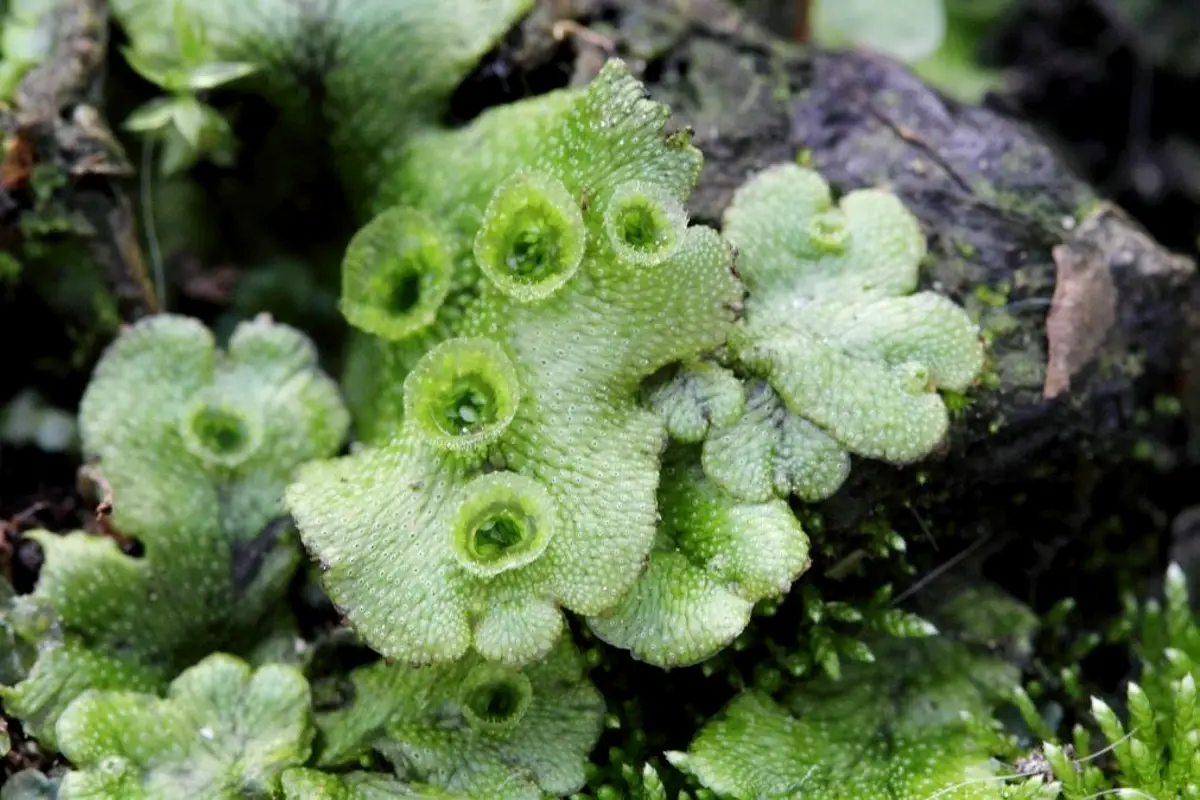
EXTERNAL MORPHOLOGY OF THALLUS OF MARCHANTIA
- Plants are thalloid, dorsi-ventral and prostrate.
- Thallus is dichotomously branched and the apex of each branch is notched.
- The dorsal side has a conspicuous midrib and many polygonal areas. These represent the underlying air chambers, each of which opens by a central air pore.
- Each air pore is compound being made of 4-S superimposed tiers of 3-4 cells each.
- Certain cup-like structures present along the midrib are known as gemma cups. These contain gemmae, the vegetative reproductive bodies.
- The ventral surface bears scales and rhizoids along the midrib.
- Scales are arranged in two to four rows on either side of the midrib. Scales are of two types-
- The simple or ligulate
- The appendiculate. The appendiculate scales have a sub-rotund appendage at their tips.
- The rhizoids are of two types
- smooth walled
- The inner wall of the smooth walled rhizoid is smooth, while that of the tuberculate rhizoid has tuber-like or peg-like ingrowths. These appear just like circular dots in surface view.
- The genus is dioecious, male and female thalli being different.
- The sex organs are present on the stalked male and female receptacles. The male receptacle is known as antheridiophore and the female as archegoniophore. These structures arise from the growing apices of the thallus.
STUDY OF ANATOMY OF THALLUS OF MARCHANTIA
- Thallus is dorsiventrally differentiated into an upper photosynthetic or assirrulatory region and a lower storage region.
- Photosynthetic region is differentiated into upper epidemis and air chambers.
- Upper epidermis is interrupted by compound, barrel-shaped air pores which open below into air chambers.
- Air pore is made of 4-S superimposed tiers of cells.
- Each air chamber is filled with many branched assimilatory or photosynthetic filaments. The cell of these filaments and epidermis possess many chloroplasts.
- Storage region is thick in the centre and gradually narrows towards margins.
- Storage region consists of compactly arranged parenchymatous cells. A few cells are filled with oil bodies and mucilage. S. The cells in the midrib or centre are slightly thickened to serve for conduction.
- The lower surface of the thallus is bound by the lower epidermis, which bears scales (two types) and rhizoids (two types) in the middle region.
STUDY OF VEGETATIVE REPRODUCTIVE STRUCTURE : THE GEMMA CUP
- Outline is goblet-shaped with an outer wall and central cavity.
- The outer wall shows outer photosynthetic region and inner storage region.
- The internal structure of photosynthetic region and storage region is similar to that of thallus.
- From the floor of the central cavity arise numerous discoid gemmae.
- Intermingled with gemmae are many mucilage hairs or cells.
- The gemma cup arises as a part of the thallus. It remains attached with the thallus by its base.
- Gemmae is one-celled, stalked structure. The stalk keeps gemma attached to the base of the gemma cup.
- The disciform gemma has two shallow notches on both the lateral sides. Each notch possesses a row of apical cells.
- Towards the periphery of the gemma colourless oil cells are present. Inner to them are the rhizoidal cells.
- All the cells of gemma except the oil cells and rhizoidal cells contain chloroplast.
STUDY OF STALK OF THE RECEPTACLE OF MARCHANTIA
- The stalk is dorsi ventrally symmetrical.
- It shows 2 rhizoidal grooves on the lower side, situated one on either side. It contains two types of rhizoids.
- Upper side has photosynthetic region divided into many air chambers. It is similar to photosynthetic region of the thallus.
- Stalks of both male and female receptacles are similar in structure.
MALE SEX ORGANS OF MARCHANTIA
- The antheridiophore consists of 0.5 to 2.0 cms long stalk, bearing at its apex one eight lobed disc.
- The peltate disc is slightly convex. The internal structure resembles with that of the thallus.
- Epidermis is interrupted below by barrel-shaped air pores, each opening below, into an air chamber with branched assimilatory filaments.
- Alternating with air chambers, are antheridial cavities. Each antheridial cavity, that opens by an antheridial pore, has a single globular antheridium.
- The antheridia are acropetally arranged i.e. oldest is nearest the centre and youngest nearest the margins.
- It has a multicellular stalk attached to the base of the antheridial cavity.
- The globular body has a single sterile jacket layer. Many androcytes or antherozoids occupy the space inside the jacket.
FEMALE SEX ORGANS OF MARCHANTIA
- It is a stalked structure, (stalk 1 to 5 cms long) possessing a nine-rayed stellate disc at the apex. Groups of archegonia are found in between the rays. In each archegonial group, the archegonia are borne in radial rows.
- After fertilization, sporophyte is formed in the same archegonium.
- The peltate disc is convex. The internal structure is similar to that of thallus.
- Outermost is the epidermis, interrupted by air pores. These open into air chambers with branched photosynthetic filaments.
- In a young receptacle, archegonia are acropetally arranged on the upper side of the disc.
- Due to the growth in the centre of the disc (which happens only after fertilization).
- The nearly mature archegonium has swollen’ venter and a long rieck.
- The venter encloses an egg cell and a venter canal cell, while the neck has 4-8 neck canal cells surrounded by six vertical rows of jacket cells.
- The cover cells are not much distinct.
- After fertilization perianth and involucre are developed.
STUDY OF SPOROPHYTE OF MARCHANTIA
- Sporophyte develops in the same place as archegonium after its fertilization. Therefore, capsules are seen in a disc of mature archegoniophore, on the lower side. Only one sporophyte develops in one involucre.
- The sporophyte is enclosed by three coverings
- calyptra
- perigynium (perianth)
- perichaetium (involucre). It is differentiated into a foot, seta and capsule.
- Foot is basal and bulbous. Seta is middle and short and the capsule is spherical, occupying the distal end of the sporophyte.
- Capsule has a single layered jacket, inside which lie many spores and elaters. Spores are arranged in tetrahedral tetrads.
- A spore has an outer thick sculptured exine and a thin uniform intine. Every spore is uninucleate with rich cytoplasm.
- The spores are very small in size.
- Elaters are spindle-shaped and each possesses 2 spiral thickening bands. These are hygroscopic and help in the dispersal of spores.
IDENTIFICATION
- DIVISION – Bryophyta
- True roots absent.
- True vascular strands absent.
- Class:- Hepaticopsida
- Mostly thalloid.
- Rhizoids without septa.
- Chloroplasts without pyrenoids.
- No columella in capsule.
- Order– Marchantiales
- Scales present.
- Two types of rhizoids present
- Air chambers and air pores present.
- Family – Marchantiaceae
- Sex organs borne on stalked receptacles.
- Air pores barrel-shaped.
- Elaters in capsule
- Genus – Marchantia
- Assimilatory filaments branched.
- Scales ligulate and appendiculate both.
- Gemma cup not crescent-shaped.
REFERENCES
Leave a Reply