Contents
- 1 CLASSIFICATION OF FUNARIA (CORD MOSS)
- 2 EXTERNAL FEATURES OF GAMETOPHYTE OF FUNARIA (CORD MOSS)
- 3 ANATOMY OF AXIS OF FUNARIA (CORD MOSS)
- 4 ANATOMY OF LEAF
- 5 STUDY OF GEMMAE
- 6 ANTHERIDIAL BRANCH AND ANTHERIDIUM
- 7 Archegonial branch and archegonium
- 8 EXTERNAL FEATURES OF SPOROPHYTE
- 9 INTERNAL STRUCTURE OF SPOROPHYTE
- 10 PERISTOME
- 11 IDENTIFICATION
- 12 REFERENCES
CLASSIFICATION OF FUNARIA (CORD MOSS)
Kingdom :- Plantae
Division :- Bryophyta
Class :- Bryopsida
Sub Class :- Bryidae
Order :- Funariales
Family :- Funariaceae
Genus :- Funaria
Funaria is very common in hills as well as in plains. It is generally found growing on moist walls and tree trunks, during rainy season.
EXTERNAL FEATURES OF GAMETOPHYTE OF FUNARIA (CORD MOSS)
- The gametophyte shows a prostrate underground protonema and an erect leafy gametophore.
- The gametophore that arises from protonema is differentiated into
- rhizoids
- axis or ‘stem’
- leaves
- Many rhizoids are present at the base. These are slender, branched, and multicellular. The septa are oblique.
- Young rhizoids are colourless while mature are coloured brown. They also develop chloroplast and become green if exposed to sunlight.
- The axis is erect and branched. It is 1-3 cms high. The branches arise below a leaf and are thus extra-axillary
- The stem and branches are covered with small, simple, sessile and spirally arranged leaves with 3/8 phyllotaxy.
- The leaves at the apex of the gametophore are crowded to form a bud-like head.
- Each leaf is nearly ovate in shape and bears a clear midrib except when young.
- Sex organs are borne at the apices of the axis.
ANATOMY OF AXIS OF FUNARIA (CORD MOSS)
- The transverse section shows an almost circular outline.
- It is differentiated into
- an epidermis.
- cortex.
- the central cylinder.
- The single-layered epidermis, with tangentially elongated cells has chloroplasts and bounds the underlying cortex.
- The multilayered cortex surrounds central cylinder. Peripheral cells of the mature cortex are slightly thick walled than the rest.
- Near the periphery of the cortex, small leaf traces with blind ends are present.
- The outer cells of the cortex sometimes contain the chloroplasts.
- The central cylinder is present in the centre. The cells are vertically elongated, smaller and the walls are slightly thickened. The cells are dead due to lack of protoplasm. The cylinder takes part in the conduction of water and food materials.
ANATOMY OF LEAF
- The leaf consists of a single layer of cells containing chloroplasts except in the middle where it forms a distinct midrib.
- The center of the midrib is occupied by a small strand of narrow and slightly thick-walled cells.
STUDY OF GEMMAE
- These are vegetative reproductive structures.
- Multicellular and green gemmae are produced on stem and leaves. On detachment, these germinate to give rise to new plants.
- Gemmae, when grow on rhizoids, become brown in colour and are then known as bulbils.
- Each gemma is composed of 8-12 cells. It is transversely and vertically septate.
ANTHERIDIAL BRANCH AND ANTHERIDIUM
- The sex organs are present at the apices of branches. These are enclosed by a/ group of leaves at the apex.
- At the tip of the stem, is an antheridial branch or ‘male flower’ -a cluster of antheridia.
- Intermingled with antheridia are multicellular capitate hairs, known as paraphyses.
- Both antheridia and paraphyses are surrounded by large leaves, known as perichaetial leaves.
- In the antheridial branch antheridia in various stages of development occur together.
- The mature antheridium consists of massive stalk and a club-shaped body.
- The body has a single layered outer jacket, the cells of which contain chloroplasts.
- At the apex of the jacket is an operculum, which helps in liberation of antherozoids.
- A dense central mass of androcytes lies within the jacket.
Archegonial branch and archegonium
- The sex organs are situated at the apices of branches inside the cluster of leaves.
- The archegonia also arise in clusters at the apex of the archegonial branch.
- Intermingled with archegonia are paraphyses.
- The archegonia and paraphyses are surrounded by closely folding, unmodified leaves.
- All the archegonia of this cluster are almost of the same age and developmental stage.
- The nearly mature archegonium is a multicellular, stalked structure, with a broad venter and narrow twisted neck.
- The wall of the venter is double layered. The neck consists of six longitudinal rows of cells surrounding a central canal.
- In the neck there are six or more neck canal cells and the venter has one venter canal cell and one egg cell.
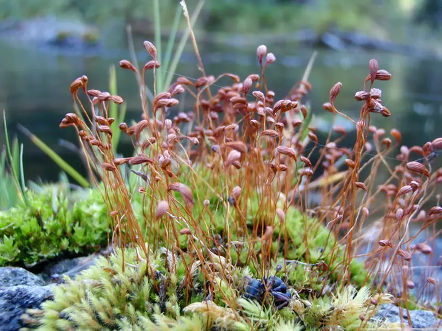
EXTERNAL FEATURES OF SPOROPHYTE
- A gametophyte shows a sporophyte attached to it.
- The sporophyte is developed at the apex of the archegonial branch.
- A mature sporophyte shows three parts (i) foot, (ii) seta and (iii) capsule.
- Foot is poorly developed and is embedded in the apex of the archegonial branch.
- Seta is long, slender and twisted. It bears a capsule at the top.
- The capsule is slightly oblique and pear-shaped. Calyptra covers the apex of capsule.
INTERNAL STRUCTURE OF SPOROPHYTE
- s. of the capsule can be divided into three regions-(i) apophysis, (ii) theca proper and (iii) upper region.
- Apophysis is the basal region. In its center is a conducting stand in continuation with that of seta.
- Around the conducting strand are few layers of cells with intercellular spaces and chloroplast. The epidermis in this region is ventilated (stomata present).
- The theca proper is the fertile region. It has a central columella, the upper part of which is cone-shaped, projecting into the concavity of the operculum. On the basal end, it is connected with the central tissue of the apophysis.
- Around the columella is a U-shaped spore sac, broken at the base, thus separating the two arms of U.
- Spore sac has an outer wall of 3-4 layers of cells and an inner of one layer. Between these, only spores are present, elaters being absent.
- Each spore has an inner hyaline endosporium and a coloured, almost smooth exosporium.
- Inside the endosporium is the cytoplasm, with a nucleus, oil globules and chloroplasts.
- Outside the spore sac, is an air space that is divided into many air cavities by green filaments which run from the external tissue of the wall to the outer wall of the spore sac.
- The wall of the capsule is many layered. Two to three inner wall layers of the capsule in theca region are green and show intercellular spaces while outermost 2-3 layers just beneath the epidermis are compact parenchymatous and colourless.
- The upper region consists of operculum and peristome. It is marked off by a conspicuous constriction, immediately below which is a rim and above the annulus.
- Calyptra covers the capsule. The peristome teeth encircle the operculum.
PERISTOME
- The peristome consists of 2 rows of curved triangular plate-like teeth. Each row has 16 teeth.
- Outer peristomial teeth are ornamented with thick transverse bands and are spirally twisted to the left.
- Outer and inner peristome.
- Inner peristomial teeth are colourless, shorter and comparatively more delicate.
- The bases of inner peristome teeth are directly covered by the teeth of the outer peristome, but as they move away from the base, they curve, thus narrowing the slits between outer peristome teeth.
- Hygroscopic movements in the outer peristome teeth assist in liberation of spores from capsule.
IDENTIFICATION
- DIVISION – Bryophyta
- True roots absent and instead are present the rhizoids.
- No true vascular strands.
- Class:- Bryopsida
- Gametophore erect and leafy,
- Rhizoids multicellular with oblique septa.
- Sub class :- Bryidae
- Leaves with distinct midrib.
- Seta long.
- Spore sac usually separated from the capsule wall by air space
- Order– Funariales
- Leaves ovate or spathulate.
- Peristome usually double
- Calyptra usually distended.
- Family – Funariaceae
- Calyptra has a long beak.
- Capsule pyriform and somewhat dropping.
- Genus – Funaria
- Leaves arranged spirally and 3/8 phyllotaxy.
- Stem internally distinguished into an epidermis, cortex and conducting strand.
- Leaves crowded at the apex to form a bud-like head
REFERENCES
Leave a Reply