Contents
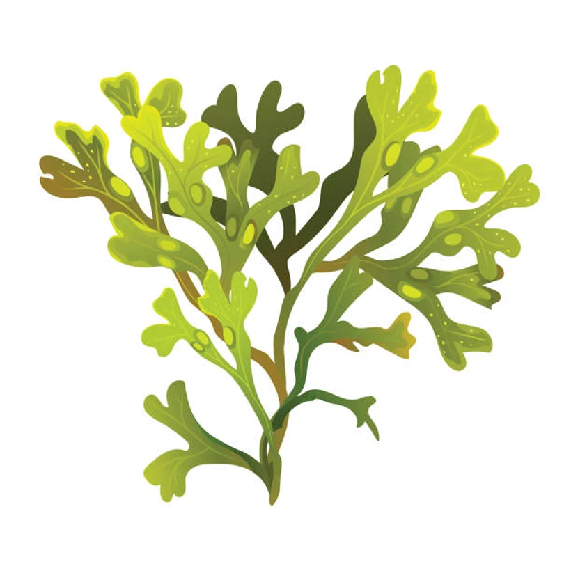
FUCUS
CLASSIFICATION OF FUCUS
Sub-division :- Algae
Class :- Phaeophyceae
Order :- Fucales
Family :- Fucaceae
Genus :- Fucus
Alga is exclusively marine. It is found attached to the rocks in the intertidal rocky coasts of the colder seas of the northern hemisphere. It is abundantly found along the coasts of British Isles, Northern European countries and Atlantic coast of America. During summers, rocks exposed to low tide remain exclusively covered with Fucus.
EXTERNAL FEATURES OF THALLUS OF FUCUS
- Thallus is flat and dichotomously branched.
- It is attached by a rounded disc-shaped holdfast.
- A mid-rib stands erect from the holdfast. It is prominent in older parts of the thallus than in younger regions.
- Thallus bears many flat strap-like, dichotomously branched blades or wings with smooth or entire margins.
- Some species of Fucus bear air bladders within the thallus. These regions of the thalli appear inflated.
- Wings show small openings of sterile conceptacles (also known as cryptostomata or cryptoblasts.
- Fertile conceptacles are terminal. These swollen parts which lack midrib are called receptacles.

INTERNAL STRUCTURE OF WING OF FUCUS
- Mature wing is internally differentiated into
- meristoderm,
- cortex
- medulla.
- The meristoderm. It is enveloped by thin cuticle consisting of small rectangular and closely compacted cells filled with dark brown plastids. This colour is due to a carotenefucoxanthin (chlorophyll in these cells can be demonstrated by immersing a portion of the wing in freshwater. This dissolves fucoxanthin and chlorophyll is visible).
- Cortex is composed of relatively large cells, size of which gradually increases toward the center. The number of plastids in the cells gradually decreases towards medulla.
- Medulla is located centrally. Cells are narrow, filamentous and loosely arranged. Spaces between the cells are filled with mucilage.
- Along with medullary cells, running longitudinally are elongated cells-hyphae (which give mechanical support to the thallus).

INTERNAL STRUCTURE OF MID RIB OF FUCUS
- Tissues are differentiated into
- outermost meristoderm,
- middle cortex
- innermost medulla.
- Meristoderm consists of small, rectangular and compacted cells arranged in a single layer. It is covered with a thin cuticle. The cells are rich in chromatophores.
- The cortex forms a wide zone. The cells near the periphery are closely compacted but become loose and larger in size towards the center
- Medulla shows irregular and longitudinal arrangement of hyphae, their narrow and thick nature making a compact and solid central tissue.

STRUCTURE OF MALE CONCEPTACLE OF FUCUS
- Plants are either monoecious or dioecious.
- The swollen tips of the branches are called receptacles.
- Each receptacle has many cavities, each being known as conceptacle.
- In monoecious species, antheridia and oogonia are present either in the same conceptacle or in two different conceptacles of the same plant.
- In dioecious species antheridia and oogonia bearing conceptacles are found on male and female plants respectively.
- T.s. of male conceptacle shows a flask-shaped cavity opening by a pore, called ostiole. 7
- A few hair-like periphyses project outside from ostiole.
- The wall of conceptacle is continuous with the external layer.
- The floor of the cavity bears multicellular hairlike paraphyses and antheridia.
- Antheridia are located on highly branched antheridial hairs or on short hairs arising from the wall of the conceptacle.
- Antheridium is stalked, unicellular, oval with double layered wall.
- Antheridium produces many pear-shaped, uninucleate and biflagellate antherozoids.

STRUCTURE OF FEMALE CONCEPTACLE OF FUCUS
- Plants are either monoecious or dioecious.
- The swollen tips of the branches are called receptacles.
- Each receptacle has many female conceptacles.
- In monoecious species antheridia and oogonia are present either in the same conceptacle or in two different conceptacles on the same plant.
- In dioecious species, antheridia and oogonia bearing conceptacles occur on male and female plants respectively.
- T.s. of female conceptacle shows a flask shaped structure, the cavity of which opens by a pore called ostiole.
- A few hair-like structures called periphyses project from near the ostiole.
- The wall of the conceptacle is continuous with the external layer.
- The floor of the cavity bears many multicellular and branched paraphyses and oogonia.
- Oogonia arise singly, directly from the wall of the conceptacle.
- Oogonium is shortly stalked, globose, swollen and has three layered thick wall.
- Mature oogonium shows eight oospheres or eggs.

IDENTIFICATION OF FUCUS
- Sub-division– Algae
- Presence of a simple thallus.
- Chlorophyll present
- Cell wall made of cellulose.
- Class – Phaeopbyceae
- Yellowish-brown chromatophores.
- Photosynthetic reserve-Iaminarin and mannitol.
- Motile reproductive cell-pyriform and flagellated
- Flagella laterally inserted and unequal.
- Order – Fucales
- Plants parenchymatous with complex morphological and anatomical differentiation
- Medulla filamentous
- Absence of asexual reproduction
- Sex organs in conceptacles.
- Family – Fucaceae
- Axes subterete to alate with mid-rib, but not foliar.
- Vesicles if present intercalary.
- Oogonia with eight oospheres.
- Genus – Fucus
- Plants erect.
- Holdfast disciform or irregular.
- Branching dichotomous or sub-pinnate.
- Branches strap-shaped with a more or less distinct mid-rib.

REFERENCES :-
Leave a Reply