Contents
- 1 CLASSIFICATION OF EQUISETUM (HORSE TAILS MOSS)
- 2 EXTERNAL FEATURES OF EQUISETUM (HORSE TAILS MOSS)
- 3 ANATOMY OF ROOT OF EQUISETUM (HORSE TAILS)
- 4 ANATOMY OF INTERNODE OF AERIAL SHOOT OF EQUISETUM
- 5 ANATOMY OF NODE OF AERIAL SHOOT OF EQUISETUM
- 6 ANATOMY OF RHIZOME OF EQUISETUM
- 7 SPORE PRODUCING ORGANS :- L.S. CONE OF EQUISETUM
- 8 SPORE PRODUCING ORGANS :- T.S. CONE OF EQUISETUM
- 9
- 10 STUDY OF SPORES OF EQUISETUM
- 11
- 12 STUDY OF PROTHALLUS OF EQUISETUM
- 13 IDENTIFICATION OF EQUISETUM
CLASSIFICATION OF EQUISETUM (HORSE TAILS MOSS)
Kingdom :- Plantae
Division :- Pteridophyta
Sub-division :- Sphenopsida
Class :- Polypodiopsida
Order :- Equisetales
Family :- Equisetaceae
Genus :- Equisetum
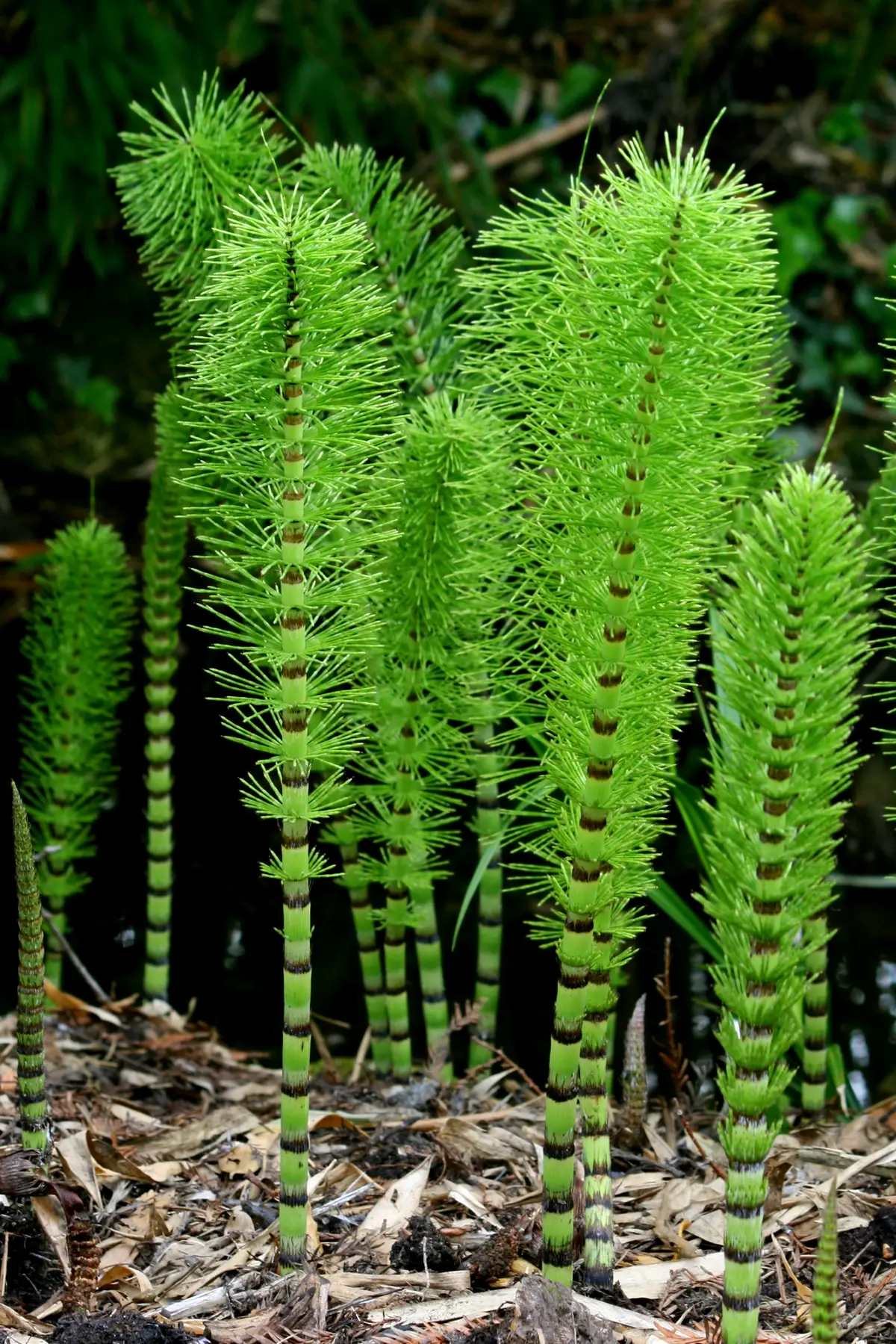
EXTERNAL FEATURES OF EQUISETUM (HORSE TAILS MOSS)
- The plants are erect and bushy.
- The plant is differentiated into roots, rhizome, aerial branches and leaves.
- The underground rhizomehas distinct nodes and internodes. The nodes bear aerial branches and roots.
- The rootsare produced on the lower side of the node. These are slender and fibrous.
- The aerial stemsare less than a metre in height with characteristic joints. Stem is rough due to the deposition of silica.
- The aerial branchesfall into two general categories-
- typical sterile branches which are green and branched, and
- typical fertile branches which are non-green, unbranched and terminate in a cone. Such branches die after the spores are shed.
- Some species have green, branched fertile shoots, with a cone at the apex of each lateral branch. Such branches do not die after the spores are shed.
- Organization of the rhizome and aerial branch is the same, but is best seen in aerial branches.
- Each internode of an aerial branch is longitudinally ribbed. The number of ridges is same as the number of leaves, and each leaf stands directly above a ridge present in the internodes below.
- The ridges on the stem of successive internodes alternate, as also the leaves of the successive nodes.
- Leaves are simple, small, scaly, whorled and fused laterally and possess longer or shorter free tips.
- Leaves are present at nodes in whorls. Each whorl forms a sheath closely appressed to the node. The number of leaves in a whorl varies with the species and the size of the stem.
- The leaves are non-chlorophyllous and scaly. These alternate at the successive nodes.
- The branches develop at the node in between each two leaves. Therefore, the branches are equal in number to the leaves and appear to arise in a whorl.
ANATOMY OF ROOT OF EQUISETUM (HORSE TAILS)
- The section appears almost circular in outline.
- Epidermisis single layered and possesses a few root hairs.
- The cortexis often divided into an outer cortex and an inner cortex.
- The outer cortexis a few layered deep. It is made of thick walled cells.
- The inner cortexis also a few layered deep. The cells are large sized and parenchymatous with intercellular spaces.
- Endodermisseparates from the vascular tissues. It is two layered-outer and inner endodermis. The pericycle is absent.
- The vascular bundleshows a single, large metaxylem element in the centre with 3 to 4 protoxylem, triarch to tetrarch elements surrounding it. The number of protoxylem groups increases with increase in the diameter of the root.
- The angles between the protoxylem are occupied by phloem.
ANATOMY OF INTERNODE OF AERIAL SHOOT OF EQUISETUM
- The outline is wavy with ridges and grooves.
- The tissues are organised into epidermis, cortex, stele and a pith cavity.
- The epidermisis cuticularized with tangentially elongated and silicified cells.
- The stomataare mostly found in the grooves. The guard cells are surrounded by two subsidiary cells, one on either side.
- Cortexfollows the epidermis and is highly differentiated. It is divided into outer and inner cortex.
- Outer cortex,below the ridges has a group of sclerenchyma. Small patches of sclerenchyma may also occur, below the grooves.
- Beneath the ridges radially elongated chlorenchymatous cells (palisade tissue) are present. The amount of palisade beneath the grooves is lesser.
- The inner cortexis composed of large and thin-walled, parenchymatous cells.
- Vallecular canals are present in the cortex. These are situated below the grooves.
- The stele is an ectophloic siphonostele that consists of ring of vascular bundles.
- Endodermisoccurs at different positions in different species.
- Most commonly, the endodermis forms a simple sheath, outside the ring of bundles.
- In some cases, in addition, there is also an internal endodermis and outer endodermis dips in between the bundles.
- In third condition, each bundle is surrounded by an individual endodermis.
- Pericyclelies below the endodermis.
- The vascular bundlesare collateral and endarch, arranged in a ring and each bundle lies below each ridge.
- Each bundle has one inner strand of protoxylem and two outer of metaxylem.
- The protoxylem elements lie on the sides of a protoxylem lacuna, the carinal canal, formed by the disintegration of protoxylem elements.
- The two metaxylem groups lie on two lateral sides of carinal canal (Le. on the shoulders of the bundle).
- The rest of the tissue of the vascular strands is parenchymatous.
- Pith cavity known as central canal lies in centre.
Features of special interest :-
Anatomy shows both xerophytic as well as hydrophytic characters.
Xerophytic characters :-
- Presence of ridges and grooves.
- Position of stomata in grooves.
- Thick cuticle over epidermis.
- Well developed sclerenchyma below the ridges.
- Presence of palisade.
Hydrophytic characters :- Presence of vallecular, carinal and central canals.
ANATOMY OF NODE OF AERIAL SHOOT OF EQUISETUM
- The section shows distinct ridges and grooves.
- The anatomy is almost similar to that of internode except for a few differences.
- The section shows epidermis, cortex, stele and nodal diaphragm instead of pith cavity in internode.
- The epidermisis the outermost thickly cuticularised layer.
- The cortexis divisible into outer, middle and inner cortex.
- The outer cortexis sclerenchymatous. It is followed by middle cortex made of palisade (chlorenchyma) tissues.
- The inner cortexis parenchymatous and occupies most part of the section.
- Vallecular canalsare absent. Many leaf traces and branch traces are found scattered all over the inner cortex.
- Vascular bundles :-Instead of ring there is a complete vascular cylinder with outer ring of phloem enclosing a ring of xylem.
- Leaf and branchtraces are given off from the vascular cylinder. Leaf traces arise beneath the ridges and do not produce leaf gaps in the vascular cylinder. Branch traces arise beneath. the grooves.
- Nodal diaphragm :-In the centre there is parenchymatous or sclerenchymatous tissue. It is known as nodal diaphragm. In L.s. it appears like an arc. The internodes easily break and separate at these places.
ANATOMY OF RHIZOME OF EQUISETUM
- The outline is wavy with ridges and grooves.
- Epidermis :-This is the outermost thickly cuticularised layer. Stomata are absent.
- The cortexconsists of a few layers of sclerenchyma just below the epidermis and a large zone of parenchyma spread upto the ring of vascular bundles.
- Large vallecular canalsare present in the parenchymatous cortex below the grooves.
- Endodermisis single layered and encloses a ring of vascular bundles.
- Each bundleis located below the ridge.
- The bundle is conjoint, collateral and endarch.
- The bundle has a large protoxylem lacuna, carinal canal.
- Pith cavity :-The centre has a large cavity, called pith cavity
SPORE PRODUCING ORGANS :- L.S. CONE OF EQUISETUM
- s. of the cone shows cone axis and attached sporangiophores.
- Cone axis is centrally located.
- It bears sporangiophores in whorls which are mostly alternate though not regularly.
- At the base of the cone is a calyx-like whorl, the annulus (which most probably represents a modified leaf whorl).
- The sporangiophores are attached to the cone axis at right angles with its stalk.
- The stalk holds a polypogonal peltate disc at right angles to it. The peltate discs of sporangiophores fit closely to form a protective cover for the sporangia below.
- Sporangia appear attached on the lower side of the disc.
- Each sporangium is elongated and sac-like. It has one-layered jacket that encloses numerous spores.
SPORE PRODUCING ORGANS :- T.S. CONE OF EQUISETUM
- s. of cone shows a cone axis and sporangiophores attached to it.
- Centrally located part is called cone axis.
- Sporangiophores are attached in a whorl.
- Each sporangiophore consists of a stalk and a disc.
- Stalk keeps the disc attached to cone axis.
- The peltate disc bears sporangia on the underside, with one layered jacket which enclose the spores.
- Each sporangium appears elongated and cylindrical.
- Sporangiophore is one of the units, of which cone is made of.
- These are attached to the central cone axis in successive whorls.
- Each sporangiophore consists of a stalk and a polygonal peltate disc.
- The stalk is attached to the cone axis on one side and to the peltate disc on the other.
- About 5-10 cylindrical sporangia are arranged in a ring near the margins on the lower side of the disc.
- Sporangium has a one layered jacket with helical thickenings.
- Numerous spores, all similar (homosporous condition) are present in the sporangial cavity.
- A longitudinal line of dehiscence is also clearly seen.
STUDY OF SPORES OF EQUISETUM
- The spores consist of a four layered wall.
- Surrounding the two usual wall layers, there is a third cuticular layer known as the middle layer and a fourth, thick, outermost layer known as perispore.
- The peri spore of each spore is differentiated into four narrow spirally wound bands, with flatspoon tips, all attached at the common point.
- These projecting bands are called as “elaters”, but are very different from the elaters of bryophyta.
- The elaters are hygroscopic and with the changes in the atmospheric humidity, they coil and uncoil. (this can be observed under the microscope by allowing the wet spores to dry on a slide).
- Each spore in a section shows a single nucleus with rich cytoplasm and all the four wall layers.
STUDY OF PROTHALLUS OF EQUISETUM
- Both male (antheridia) and female (archegonia) sex organs are borne on the same prothallus. Thus it is monoecious.
- In younger stages only antheridia are developed. Therefore, small and younger prothalli show antheridia only. In older prothalli, however, archegonia are found. Hence it is protandrous.
- The multicellular central part gives out many flat branches. These branches further get irregularly dissected in uniseriate filaments.
- From the central region, long, brown and unbranched rhizoids are also given off.
- Archegonia remain embedded in the tissue of the prothallus, at the place where branches are given out.
IDENTIFICATION OF EQUISETUM
- DIVISION – Pteridophyta
- True roots generally present (except in Psilopsida),
- True vascular strand present.
- Sub-division:- Sphenopsida
- Stem branched, articlliated, ridged and furrowed with distinct nodes and internodes.
- Leaves microphyllous, small, scaly and in whorls at nodes.
- Order– Equisetales
- Stem branched. Branches borne in transverse whorls.
- Internodes alternate with one another.
- Vascular cylinder endarch, siphonostele.
- Family – Equisetaceae
- Sporangia borne on sporangiophores which form a compact cone
- No secondary growth.
- Genus – Equisetum
- Leaves scaly and colourless.
- Sunken stomata in grooves.
- Presence of palisade in the stem,
- Presence of valecular, carinal and central canals.
Leave a Reply