Contents
- 1 CLASSIFICATION OF EPHEDRA
- 2 EXTERNAL FEATURES OF EPHEDRA
- 3 T.S. OF YOUNG STEM OF EPHEDRA
- 4 T.S. OF STEM SHOWING SECONDARY GROWTH OF EPHEDRA
- 5 L.S. OF WOOD. OF EPHEDRA
- 6 T.L.S. WOOD OF EPHEDRA
- 7 MALE STROBILUS OF EPHEDRA
- 8 MALE FLOWER OF EPHEDRA
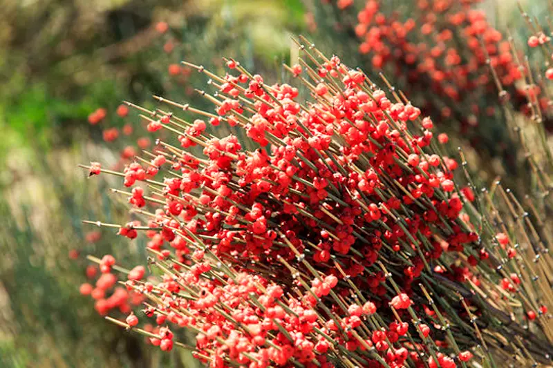
- 9 FEMALE STROBILUS OF EPHEDRA
- 10 L.S. OVULE OF EPHEDRA
- 11 L.S. SEED OF EPHEDRA
- 12 IDENTIFICATION OF EPHEDRA
- 13 REFERENCES
CLASSIFICATION OF EPHEDRA
Kingdom :- Plantae
Division :- Gymnosperm
Class :- Gnetopsida
Order :- Gnetales
Family :- Ephedraceae
Genus :- Ephedra
EXTERNAL FEATURES OF EPHEDRA
- Plants are small, bushy, trailing or climbing shrubs attaining a height of not more than 2 meters. However, E. antisiphilitica is a small tree, reaching a height of 3-5 meters.
- The plant body is branched and posseses only minute leaves at the nodes. It therefore, resembles superficially with the species of Psilotum and Equisetum.
- It is differentiated into stem, leaves and underground roots.
- The stem remains anchored by a deep tap root and many adventitious roots.
- The stem is delicate, slender and green when young. It is ribbed irregularly and is differentiated into short nodes and long internodes.
- Two or three branches arranged in whorls arise from the nodes in the axils of leaves. The branches are shed off during dry season.
- Older part of the stem may bear many branches. It becomes hard and woody due to secondary growth.
- Leaves are borne in a whorl of 2-4 at each node.
- Leaves that are small and scale-like are connate at the base and thus form a sheath around the node.
- Each scale leaf is traversed by two parallel and unbranched veins.
- Foliage leaves are completely absent.
- The male and female reproductive organs are borne in small strobili.
- The plants are mostly dioecious and bear only one type of reproductive organs. They may also be monoecious when they bear both kinds of strobilii.
T.S. OF YOUNG STEM OF EPHEDRA
- The outline of the section shows ribbed nature of the stem.
- The tissues are differentiated into epidermis, cortex and stem.
- Epidermisis the outermost layer. It is very thick and heavily cuticularized.
- The stomataare sunken. These interrupt epidermis frequently and occupy a position just on the slopes of the ridges.
- Hypodermisis sclerenchymatous and occur in small groups below the ridges.
- Cortex :-Rest of the cortical tissue is cholrenchymatous. The cells are often radially elongated and contain abundant chloroplasts. Large intercellular spaces are present between these cells.
- A few patches of sclerenchyma occur dispersed in the cortex (specially in young axis rendering hardness and resistance).
- Steleis ectophloic siphonostele. It is composed of many vascular bundles, their number being variable.
- Endodermisis single layered and is followed by a peri cycle.
- A few vascular bundlesare arranged in a ring. Each is conjoint, collateral, endarch and open.
- External phloemgroup is separated from internal xylem group by a narrow layer of cambium.
- Pithis parenchymatous and occurs in the central region.
- Nodal diaphragm :-The characteristic anatomical feature diaphragm-like plate each internode or is the presence of of cells at the base of at the node (nodal diaphragm). This helps the plant to shed off the branches at the nodes.
T.S. OF STEM SHOWING SECONDARY GROWTH OF EPHEDRA
- The section shows epidermis, cortex and primary and secondary vascular tissues.
- The epidermis and cortexremain unchanged. However, after (3-4 Years) of growth, cork develops just outside the phloem and outer tissues (epidermis, cortex, etc.) are, therefore, cast off.
- Sclerotic cells(stone cells) develop just above the zone of secondary tissue.
- Primary phloemoccurs as obliterated patches.
- Secondary phloemfonns a zone below. Phloem is composed of sieve tubes and phloem parenchyma.
- Annual ringsare distinct comprising autumn and spring wood each. These are fonned in the secondary xylem (wood).
- The secondary xylemshows a thin walled spring wood and thick walled autumn wood, successively formed in alternating zones.
- Antumn wood is made of smaller cells, while those of spring wood are bigger in size.
- Thetracheidal cells of the secondary wood are associated with broad vessels. Though absence of vessels is characteristics of the Gymnospenn, Ephedra, (i.e. order Gnetlaes) itself is an exception.
- Vesselsare most abundant in the spring wood’ and a few or none at all in the autumn wood. Spring wood is often ring porous.
- Tracheids and vesselshave uniseriatey or irregularly distributed bordered pits. Protoxylem elements of primary xylem show spiral, annular or reticulate tracheids.
- Medullary raystraverse the wood. Primary medullary rays run from primary xylem to primary phloem while secondary medullary rays run from secondary xylem to secondary phloem.
- Medullary rays are uniseriate in the young stem but are very broad and long (multiseriate) in the old stem.
- Primary xylemgroups are present at the end of the secondary wood near the pith. These are endarch.
- Pithis large and parenchymatous. It occupies the centre.
Features of special interest
It shows the following xerophytic characters.
- Thickly cuticularized epidermis.
- Sunken stomata.
- Palisade and spongy parenchyma in the cortex.
- Patches of sclerenchyma.
- Shedding of branches.
- Presence of nodal diaphragm.
- Vessel in the secondary wood.
L.S. OF WOOD. OF EPHEDRA
- It shows the presence of secondary xylem and medullary rays.
- Secondary xylem consists of tracheids with bordered pits on their radial walls.
- The bordered pits are circular or slightly elliptical. These may form reticulations (mostly due to the dissolution of walls of cavities of the pits) and such perforations, being characteristic of Ephedra, are known as Ephedroid perforations. Bordered pits are either scattered or arranged in 2 or 3 tight rows. (They are never polygonal due to mutual compression).
- Special cellulose thickening – Bars of Sanio are also present below the pits.
- A few vessels present show bordered pits which are scattered or may remain arranged in 2 or 3 rows. The apertures of the bordered pits are commonly horizontally oriented. End walls are also perforated.
- Medulllary rsays are uniseriate or muItiseriate. These run horizontally. In this plane medullary rays are cut lengthwise and their length and height can be observed.
- Medullary rays may range up to 40-50 cells in height.
- Each medullary ray in the region of secondary xylem is composed of ray cells and ray tracheids dispersed regularly.
- Ray cells are thick walled as well as thin walled. These occur in the same medullary ray. Their tangential walls possess bordered pits or slit-like openings.
- Ray tracheids are thick walled. Their radial and tangential walls are pitted, pits being bordered.
- In the region of phloem, medullary ray is made of starch cells surrounded by albuminous cells on both sides.
T.L.S. WOOD OF EPHEDRA
- In this plane tracheids, vessels and medullary rays are cut transversely.
- Bordered pits are seen in surface view.
- The bordered pits show usual over-arching dome-shaped structure and a small disc-torus.
- Medullary rays are transversely cut and as such their height and breadth can be determined.
- Rays are elongate and many tangential walls show simple slit-like pits.
MALE STROBILUS OF EPHEDRA
Reproductive parts are borne in strobili. One of the following conditions may be found :
- Usually male and female strobili are different; in such a case, the strobilus may be termed as monosporangiate. These strobili may be borne on two different plants (dioecious sps.).
- Sometimes one plant may bear both the strobili (monoecious sps.).
- A few plants, sometimes, bear both the reproductive parts in one strobilus only (bisporarangiate strobilus), e.g. E. Joliata and E. intermedia. In such cases, male flowers are situated below the female flowers which occur at the higher level in the same strobilus
COMMENTS :-
- The strobilus resembles inflorescence, spike.
- Each stobilus consists of an axis which bears decussately arranged sterile scales and stamens.
- Male spike (male strobilus or staminate strobilus) arises in the axil of scale leaf.
- Each spike is generally round in shape but may be ovoid or spherical.
- A spike has a short axis with many scaly bracts. The bracts are arranged in decussate pairs. The number of pairs varies from 2-12.
- In the axil of each bract arises a single male flower.
MALE FLOWER OF EPHEDRA
- A male flower has a perianth of bract scale which encloses a stamen.
- Stamen consists of a stalk (variously termed as column or antherophore). It bears 2-5 microsporangia or anthers at its tip.
- Each microsporangium is a bilocular structure. It has two wall layers and a prominent tapetal layer which encloses pollen grains or microspores.
- Each microspore is elliptical and has an outer thick and ribbed exine and a thin intine.
- A micro sporangium opens by apical part (apical dehiscence).
FEMALE STROBILUS OF EPHEDRA
- The strobilus resembles spike inflorescence.
- Each strobilus consists of an axis bearing decussately arranged sterile bracts (scales) and ovules.
- The female or ovulate strobili arise in the axil of scale leaves. The female strobilus is also sessile and not so richly branched as the male.
- The apex of ovulate strobilus is mostly acute.
- The spike has a short axis on which about 4-7 pairs of bracts are arranged decussately.
- Lower most 1 or 2 pairs are sterile while terminal pairs bear short stalked ovules. The bracts are generally dry, winged and may be variously coloured.
- Each bract mostly encloses two ovules out of which one may be abortive.
L.S. OVULE OF EPHEDRA
- Ovule is covered by two integuments.
- Outer integument (involucre or perianth) is a cup-like structure, attached at the base of the ovule and free above.
- Inner integument is delicate, composed of two segments. It prolongs into a tubular process and comes out beyond the bracts and involucre at the time of pollination.
- Micropyle is an opening in between the integuments, in the upper region of the ovule.
- Nucellus lies below the integuments. A small pollen chamber is present just below the micropyle in the tissue of nucellus.
- Female gametophyte is a tissue situated below the pollen chamber. Two archegonia are present, just below the pollen chamber, in the female gametophyte.
- Haustorial region lies opposite the micropylar end. It is occupied by tissue filled with stored food material. It also gives out haustorial processes for the absorption of food and is known as haustorial region.
L.S. SEED OF EPHEDRA
- Outer integument encloses entire seed. It is thick walled.
- The inner integument (‘true integument’) persists at the micropylar end only.
- Nucellus forms shrivelled layer in the form of a disorganized sheath of cells. It is located inside the inner integument.
- Female gametophyte (endosperm) surrounds a big embryo which has two large cotyledons.
- Bracts adjacent to strobilus are fleshy and thick in a completp.ly mature seed. These form an additional envelope
IDENTIFICATION OF EPHEDRA
- DIVISION – Gymnosperms
- Absence of vessels.
- Ovules naked.
- Seeds attached with woody acales.
- Scales generally form a cone.
- Class:- Gnetopsida
- Wood with vessels.
- Flowers in compound strobili or ‘inflorescence’, unisexual, usually dioecious.
- Ovules surrounded by several envelops.
- Order– Gnetales
- Plants woody trees, shrubs or lianes
- Leaves opposite or whorled, simple
- Family – Ephedraceae
- Plants either shrubs or woody climbers
- Leaves scaly, foliage leaves atisent
- Nodal diaphragm present
- Stamens enclosed by bract
- Seeds covered with fleshy bracts.
- Genus – Ephedra(Single genus)
REFERENCES
- https://en.wikipedia.org/wiki/Ephedra_sinica
- https://www.shutterstock.com/search/ephedra
Leave a Reply