Contents
- 1 CLASSIFICATION OF ANTHOCEROS (HORN WORT)
- 2 EXTERNAL MORPHOLOGY OF ANTHOCEROS (HORN WORT)
- 3 ANATOMY OF THALLUS OF ANTHOCEROS (HORN WORT)
- 4 TUBER OF ANTHOCEROS (HORN WORT)
- 5 ANTHERIDIUM OF ANTHOCEROS (HORN WORT)
- 6 ARCHEGONIUM OF ANTHOCEROS (HORN WORT)
- 7 SPOROPHYTE OF ANTHOCEROS (HORN WORT)
- 8 IDENTIFICATION
CLASSIFICATION OF ANTHOCEROS (HORN WORT)
Kingdom :- Plantae
Division :- Bryophyta
Class :- Anthocerotopsida
Order :- Anthocerotales
Family :- Anthocerotaceae
Genus :- Anthoceros
Anthoceros is common in both hills and plains. Plants grow, as a rule, in moist, shady places on the sides of ditches or in moist hollows among rocks.
EXTERNAL MORPHOLOGY OF ANTHOCEROS (HORN WORT)
- Plant body is thalloid, somewhat lobed or radially dissected and generally suborbicular.
- The thallus is less often dichotomously branched and lack a definite midrib.
- The dorsal surface of the thallus is generally smooth, velvety or rough.
- The ventral surface bears smooth walled rhizoids only.
- On the ventral side, a few bluish spots are seen indicating the presence of filaments of blue green-alga (viz. Nostoc or Anabaena).
- Sex organs are situated on the dorsal side and are embedded in the tissue of the thallus.
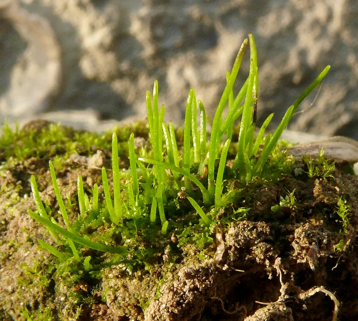
- The sporophyte, however, is linear and elongated structure, arising from the dorsal side.
ANATOMY OF THALLUS OF ANTHOCEROS (HORN WORT)
- Thallus is few cells in thickness in the middle and becomes thinner towards the margins.
- Internal structure is homogeneous (i.e. all cells are alike). Air pores and air chambers are absent.
- On upper side is present upper epidermis and lower epidermis on the lower side.
- Parenchymatous tissue lies between these two layers .
- Each parenchymatous cell has a distinct nucleus and a chloroplast.
- Each of the cells has a large chloroplast with a single pyrenoid except the cells of lower epidermis producing rhizoids. There are two chloroplasts in the cells of A. pearsonii and four in A. hallii.
- On the ventral side there are certain mucilagefilled cavities which open by slime pores, through the ventral epidermis.
- The endophytic algae Nostoc or Anabaena present in he mucilage cavities, enter through these slime pores.
- Rhizoids are smooth-walled and arise in the middle region of the thallus from the lower epidermis.
TUBER OF ANTHOCEROS (HORN WORT)
- The tubers are the vegetative reproductive structures.
- Tubers are formed under unfavourable conditions, on the dorsal side along the margins of the thallus.
- A tuber in section shows outer 2-3 corky layers, protecting the inner tissue, containing the reserve food material.
- On return of the favourable conditions, tubers develop into new thalli.
ANTHERIDIUM OF ANTHOCEROS (HORN WORT)
- Both antheridia and archegonia remain embedded in the dorsal region of the thallus and are acropetally arranged.
- Few species are monoecious (homothallic), but some are dioecious (heterothallic).
- Monoecious species are frequently protandrous (antheridia maturing first).
- The antheridia are present in the antheridial cavity or antheridial chamber with a sterile roof of 2-3 layers.
- Each antheridial cavity contains about 1-4 or more primary antheridia. Secondary antheridia arise from the stalks of primary antheridia and ultimately there may be as many as 25 in each antheridial cavity.
- The mature antheridium is a stalked, club-shaped structure with single layered jacket. Each cell possesses a prominent plastid.
- Inside the jacket, there are large number of androcytes.
ARCHEGONIUM OF ANTHOCEROS (HORN WORT)
- The thalli are generally monoecious and protandrous (antheridia maturing fust).
- The archegonia are embedded in the thallus and only the cover cells project beyond the general surface of the thallus.
- They are in direct contact with the vegetative cells, lateral to them.
- Archegonium consists of a neck and a swollen venter.
- The nearly mature archegonium has 4 cover cells, 4-6 neck canal cells, one venter canal cell and one egg cell.
SPOROPHYTE OF ANTHOCEROS (HORN WORT)
- The sporophytes are linear, 2-3 cms long, elongated structure, arising from the dorsal side of the thallus.
- The base of each sporophyte is enclosed by an involucre, made of gametophytic tissue. (The internal structure of the sporophyte can be understood by studying the transverse and longitudinal sections). s. of the sporophyte shows following characters:
- The mature sporophyte is made of a lower foot, middle meristematic zone and upper capsule.
- The foot is bulbous and is deeply rooted in the gametophytic tissue.
- The seta is absent and instead is present the meristematic zone.
- In the centre is columella, composed of 16 vertical rows of cells, extending from the base to the tip of the capsule.
- Surrounding the columella is a cylinder of sporogenous tissue which extends from the base to the tip of the capsule where it over-arches the central columella. It reveals sporogenous series, from one layered archesporium at the base, to mature spores and pseudo-elaters at the tip.
- At the base of the capsule is a single to double layered archesporium, while little higher up, archesporium is differentiated into alternately placed fertile cells (spore mother cells) and sterile cells (which form pseudo-elaters). A part of sporogenous tissue, upper to this region shows tetrahedral tetrads of spores and many pseudo-elaters. In the upper most region of the capsule are present the separated spores and pseudo-elaters.
- The wall of the capsule is 4-6 layered.
- The outermost layer forms a well defined epidermis, ventilated with stomata.
- The cells of the layers beneath the epidermis possess generally two chloroplasts each and these chloroplasts make the sporophyte partially self-sufficient with regard to food.
- Each spore is uninucleate and has a thick ornamented exospore and a thin endospore.
- Pseudo-elaters consist of 2-3 cells, joined end to end, in a simple or branched structure. (These do not possess characteristic spiral thickenings of true elaters and, therefore, are called pseudo-elaters).
IDENTIFICATION
- DIVISION – Bryophyta
- True roots absent and instead are present the rhizoids.
- No true vascular strands.
- Class:- Anthocerotopsida
- Rhizoids without septa.
- Each cell of the thallus has generally a single large chloroplast, with pyrenoid.
- Order– Anthocerotales
- thallus homogeneous
- Only smooth walled rhizoids present, scales and tuberculated
rhizoids absent.
- Family – Anthocerotaceae
- Sporophyte indeterminate in growth.
- Presence of meristematic zone
- Capsule with central columella.
- Genus – Anthoceros
- Capsule partly covered with involucre, only at the base.
- Capsule wall ventilated.
- Nostoc colonies inside the thallus
Leave a Reply