Contents
- 1 CLASSIFICATION OF ADIANTUM (MAIDEN HAIR FERN)
- 2 EXTERNAL FEATURES OF ADIANTUM (MAIDEN HAIR FERN)
- 3 ANATOMY OF ROOT OF ADIANTUM (MAIDEN HAIR FERN)
- 4 ANATOMY OF RHIZOME OF ADIANTUM (MAIDEN HAIR FERN)
- 5 ANATOMY OF RACHIS OF ADIANTUM (MAIDEN HAIR FERN)
- 6 ANATOMY OF LEAFLET OF ADIANTUM (MAIDEN HAIR FERN)
- 7 STRUCTURE OF SORUS OF ADIANTUM (MAIDEN HAIR FERN)
- 8 STRUCTURE OF A SPORANGIUM AND A SPORE OF ADIANTUM (MAIDEN HAIR FERN)
- 9 STRUCTURE OF PROTHALLUS of ADIANTUM (MAIDEN HAIR FERN)
- 10 OLD PROTHALLUS WITH SPOROPHYTE OF ADIANTUM (MAIDEN HAIR FERN)
- 11 IDENTIFICATION OF ADIANTUM (MAIDEN HAIR FERN)
CLASSIFICATION OF ADIANTUM (MAIDEN HAIR FERN)
Kingdom :- Plantae
Division :- Pteridophyta
Sub-division :- Pteropsida
Class :- Leptosporangiatae
Order :- Filicales
Family :- Adiantaceae
Genus :- Adiantum
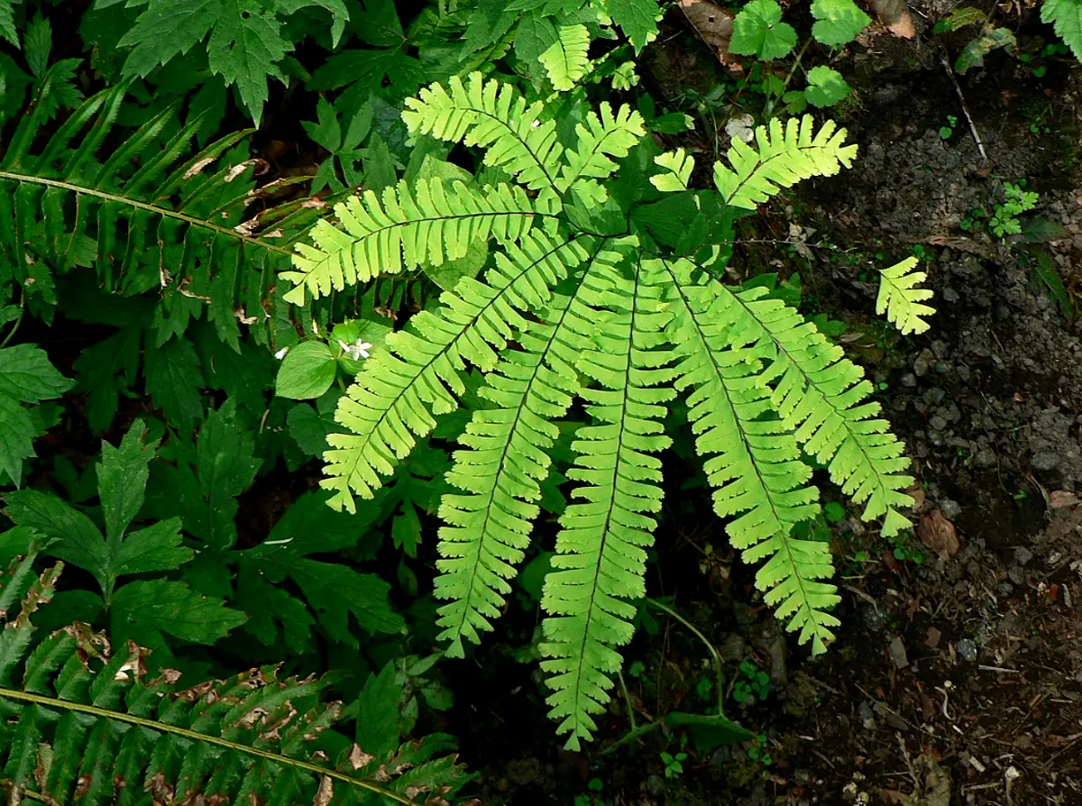
EXTERNAL FEATURES OF ADIANTUM (MAIDEN HAIR FERN)
- Plant bodyis a sporophyte. It is differentiated into roots, rhizome and leaves.
- Rootsare produced on the lower side of the rhizome. Primary root is short-lived. Secondary roots are adventitious and branched.
- Rhizomemay either be creeping or erect. It is scaly, covered with hairs and bears adventitious roots and leaves.
- Leaveswith a long petiole, are spirally or alternately arranged on the rhizome. Young leaves are circinately coiled. Young rhizome, petiole, rachis and circinately coiled leaves are covered with hairs known as ramenta.
- The leaves are compound and are borne on shining black and brittle petiole.
- Leaves are often dichotomously branched into many leaflets.
- The blade of the leaflets may be entire, and either simply or repeatedly branched. The leaflets are deltoid in shape. When fertile, the leaflet margin remains folded toward the lower side forming a false indusium which encloses many sori.
- Leafletis traversed by dichotomously branched veins which generally do not unite to form a reticulum. The venation is, therefore, of open dichotomous type.
ANATOMY OF ROOT OF ADIANTUM (MAIDEN HAIR FERN)
- The root is almost circular in outline.
- It is differentiated into epidermis, cortex and stele.
- The epidermisis single layered, cells are thin walled and tangentially elongated. It bears a few unicellular root hairs.
- Cortexis inner to the epidermis. It is multilayered and parenchymatous.
- Endodermisis single layered and is followed by a single layered pericycle.
- A protosteleis present in the centre. It is diarch and exarch.
- Xylemelements occur in the centre. Of these, metaxylem elements are present in the centre. Protoxylem groups are situated on the two opposite sides of the metaxylem (Thus xylem groups are exarch and diarch).
- Phloemsurrounds the xylem on all the sides.
ANATOMY OF RHIZOME OF ADIANTUM (MAIDEN HAIR FERN)
- Rhizome appears almost circular or guttershaped in a transection.
- It shows differentiation into epidermis, hypodermis, ground tissue and stele.
- Epidermisis single layered and bears numerous multicellular hairs.
- Hypodermisthat follows epidermis is a 2-3 layered deep and sclerenchymatous.
- Ground tissueoccupies major part of rhizome. It is parenchymatous and many layered deep.
- Steleis variable in nature, differing from one region to another. Species with elongated rhizome (A. pedatum and A. hispidulum) show actual solenostele (amphiphloic siphonostele). But commonly the stele is gutter shaped and appears as several meristeles arranged in a ring, due to numerous leaf gaps (dictyostele).
- Meristelesvarying in number, but more often 5-7, lie arranged in ground tissue in a guttershaped ring, thus exhibiting dictyostelic condition. The spaces between neighbouring meristeles are leaf gaps.
- Each of the meristeles is surrounded by a distinct, single-layered endodermis subsequently followed by a single layered pericycle.
- Xylemelements occupy the central part. Metaxylem and protoxylem are arranged in a way to form mostly mesarch condition.
- Phloemsurrounds xylem.
ANATOMY OF RACHIS OF ADIANTUM (MAIDEN HAIR FERN)
- Tissues of the rachis are differentiated into epidermis, hypodermis, cortex and stele.
- Epidermisis a single layer of cells, covered by a cuticle.
- Hypodermisfollows the epidermis. It is few layered deep. The cells are sclerenchymatous.
- Cortexforms larger part of the rachis. It is made of many layers of parenchyma.
- Steleis a protostele.
- Endodermisis single layered and is followed by a pericycle.
- The xylemgroup is almost Y -shaped. Protoxylem elements are situated at the tips of three free ends; while the rest of the part is occupied by metaxylem.
- Phloemsurrounds the xylem on all the sides.
ANATOMY OF LEAFLET OF ADIANTUM (MAIDEN HAIR FERN)
- Leaflet is differentiated into upper and lower epidermis, mesophyll, sclerenchyma and vascular bundle.
- Epidermis:- The cells of the upper and lower epidermis possess chloroplast. The lower epidermis is frequently interrupted by stomata.
- Chloroenchyma:-Just above the lower epidermis lies a single layer of compactly arranged cells containing numerous chloroplasts.
- Mesophyll:- Following this compact layer, mesophyll tissue extends up to the upper epidermis. It is undifferentiated into palisade and spongy parenchyma but is composed of loosely arranged spongy parenchyma only.
- Vascular bundle:- Each vascular bundle is surrounded by a thick sclerenchymatous sheath.
- Endodermis:- Surrounding the vascular bundle is endodermis followed by a pericycle.
- Xylem :-Centrally located xylem has protoxylem groups, facing towards the adaxial surface of the leaf.
- Phloemsurrounds xylem.
STRUCTURE OF SORUS OF ADIANTUM (MAIDEN HAIR FERN)
- The spore producing organs are the sporangia grouped in sori. Each sorus is mixed in nature and shows sporangia of different ages.
- Sori are present, along both the sides of the veins, on the dorsal side of the marginal reflexed lobe. This part remains folded towards the lower side and acts as a false indusium which is membranous and brown coloured.
- Lower part of each leaflet shows many such reflexed lobes along its margins. The reflexed part is traversed by veins in continuation with those in the unfolded part.
To study the relation of the folded part with the unfolded, cut a L.s. of leaflet in folded condition.
- It shows the upper part of the leaflet possessing vascular bundles cut longitudinally and the reflexed or folded margin on its lower side.
- This reflexed part is a portion of the leaflet and bears sporangia in sori, thus forming a false indusium.
- Many sporangia are seen arranged in groups.
- Sporangia are attached to the indusium by their long slender stalks.
To study the relation between veins, sori and sporangia with indusium, unfold the reflexed lobe and cut its T.s.
- Reflexed lobe of leaflet covers sori and is called false indusium.
- Veins traverse the indusium but do not form reticulum (open dichotomous venation).
- Along both the sides of each vein are many sporangia, attached by their long and slender stalks. This group of sporangia is called a sorus and many such sori are situated along each of the veins.
STRUCTURE OF A SPORANGIUM AND A SPORE OF ADIANTUM (MAIDEN HAIR FERN)
- Each sporangium is attached to the indusium by a slender, long and multicellular stalk.
- The sporangium (capsule) is oblong in shape and borne at the tip of the stalk.
- The wall of the sporangium is made of thin walled cells.
- Some cells of this wall lying in a vertical row are characteristically thickened on their radial and inner tangential walls. These together are called as annulus.
- On one side of the annulus are a few (2-3) thin walled cells forming a stomium wherefrom dehiscence of the sporangium takes place.
- The wall encloses many spores inside.
- Each spore is a double walled structure. The outer layer is exine which is thick and ornamented. The inner layer, called intine, is thin and smooth.
- It has a single big nucleus, surrounded by cytoplasm.
STRUCTURE OF PROTHALLUS of ADIANTUM (MAIDEN HAIR FERN)
- It is formed after the germination of a spore and is thus a gametophytic structure.
- It is leafy and heart-shaped.
- It consists of a single layer of cells, one cell in thickness, except in the central region where apical notch is situated.
- Many unicellular rhizoids are given out from the ventral surface.
- Antheridia are located in the posterior part of prothallus away from the apical notch.
- Archegonia lie near the apical notch, on the thickened, central apical cushion.
- Parts of antheridia and necks of archegonia protrude outside the general surface of the prothallus.
- All the cells of the prothallus are thin walled and bear many discoid chloroplasts.
OLD PROTHALLUS WITH SPOROPHYTE OF ADIANTUM (MAIDEN HAIR FERN)
- Sporophyte is formed as result of fertilization.
- Gametophyte (prothallus) still persists.
- Young sporophyte is differentiated into young leaves, primary root and secondary roots.
- Leaves stand erect and appear near the apical notch. They are simpler than the mature leaves. They may also be circinately coiled.
- Primary root grows on the lower side and gives out secondary roots.
IDENTIFICATION OF ADIANTUM (MAIDEN HAIR FERN)
- DIVISION – Pteridophyta
- True roots generally present (except in Psilopsida),
- True vascular strand present.
- Sub-division:- Pteropsida
- Vascular cylinder siphonostelic, with leaf gaps.
- Plants macrophyllous, leaves compound, with rachis.
- Leaves bear sporangia in sori.
- Gametophytes small, green and free living.
- Class:- Leptosporangiatae :-
- Sporangium with a jacket layer one cell in thickness.
- Definite number of spores.
- Order– Filicales
- Sori are mixed
- Family – Adiantaceae
- Sori marginal.
- Indusium oblong or linear, formed of the more of less changed and reflexed margin of the frond. opening inwardly.
- Genus – Adiantum
- Sori apparently marginal, but superficial in origin.
- Indusia globose to linear, usually many and distinct.
- Leaflet margins bearing sori are sharply reflexed.
- Open dichotomous venation of the leaflet.
Leave a Reply