Contents
CLASSIFICATION OF PELLIA
Kingdom :- Plantae
Division :- Bryophyta
Class :- Hepaticopsida
Order :- Jungermanniales
Family :- Pelliaceae
Genus :- Pellia
EXTERNAL MORPHOLOGY OF PELLIA
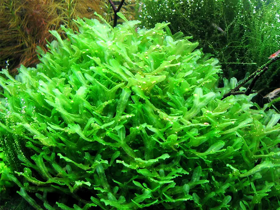
- The plants are thalloid, prostrate, drosiventral and dichotomously branched.
- On the dorsal side there is an indistinct midrib and one celled thick lateral wings, with somewhat wavy margins.
- At the apex is a notch in which growing point is situated. Club-shaped mucilaginous hairs are also present at the apex.
- The shape of the thallus depends upon moisture conditions. If the thalli grow near water, they are narrow, ribbon-like, delicate and with distinct midrib. If the thalli happen to grow on dry soil, they become shorter, thick and bear an indistinct midrib.
- Only smooth walled rhizoids are present on the ventral side towards the midrib portion. The tuberculated rhizoids and scales being altogether absent.
ANATOMY OF THALLUS OF PELLIA
- The section of the thallus shows homogeneous internal structure. Only the surface layers are little different while cells forming the rest of the tissues are all similar.
- The section shows ventral projected midrib and wings. The midrib is 8-14 cells in thickness passing gradually on either sides into one celled wings.
- The epidermal cells are smaller and contain more chloroplasts, than the other cells which have distinct starch grains and oil bodies.
- In P. epiphylla and P. neesiana there are some thick-walled cells in the midrib portion which travel longitudinally from posterior to anterior end.
- Rhizoids arise from the lower epidermis in the midrib region.
- Vegetative reproduction takes place by adventitious shoots arising either from the midrib or from margins of the dorsal side. These branch on their separation from the parent plants and grow into new plants.
MALE SEX ORGANS – THE ANTHERIDIA (PELLIA)
- Outline is goblet-shaped with an outer wall and central cavity.
- The outer wall shows outer photosynthetic region and inner storage region.
- The internal structure of photosynthetic region and storage region is similar to that of thallus.
- From the floor of the central cavity arise numerous discoid gemmae.
- Intermingled with gemmae are many mucilage hairs or cells.
- The gemma cup arises as a part of the thallus. It remains attached with the thallus by its base.
- Gemmae is one-celled, stalked structure. The stalk keeps gemma attached to the base of the gemma cup.
- The disciform gemma has two shallow notches on both the lateral sides. Each notch possesses a row of apical cells.
- Towards the periphery of the gemma colourless oil cells are present. Inner to them are the rhizoidal cells.
- All the cells of gemma except the oil cells and rhizoidal cells contain chloroplast.
FEMALE SEX ORGANS -THE ARCHEGONIA (PELLIA)
- The archegonia are found in groups of 4 to 12, just near the apical cell.
- Each archegonial group is surrounded by an involucre which may either be tubular, cylindrical or flap-like.
- Archegonia are intermingled with each other without definite arrangement of older and younger archegonia.
- A nearly mature archegonium has a short multicellular stalk, a broad venter and a long neck.
- The jacket of a neck consists of five vertical rows of cells and encloses usually 6-8 neck canal cells.
- The venter is two-layered thick and encloses a single venter canal cell and an egg cell.
- The cover cells are 4 in number but are not very much distinct.
SPOROPHYTE OF PELLIA
- The mature sporophyte consists of foot, seta and capsule.
- The foot is very prominent with its edges overlapping the basal portion of seta, thus assuming a collar-shape.
- The seta is short when young but becomes very much elongated at maturity.
- The capsule is covered by calyptra and involucre respectively.
- The capsule is nearly spherical and consists of an outer jacket composed of 2 or 3 layers.
- The jacket layers of capsule have radial thickenings except at 4 places at the top wherefrom the dehiscence takes place.
- Inside the jacket at the base of the capsule is present a sterile tissue, known as elaterophore. On this elaterophore are attached some 20 to 100 elaters, radiating into the cavity of capsule.
- In the remaining cavity of the capsule are present spores and elaters.
- The spores are arranged in tetrahedral tetrads and are formed by lobing of spore mother cell.
- Each spore is unicellular. The elater is a long, slender, spindle-shaped, structure with generally 2 but sometimes 3-4 spiral thickening bands.
IDENTIFICATION
- DIVISION – Bryophyta
- True roots absent and instead are present the rhizoids.
- No true vascular strands.
- Class:- Hepaticopsida
- Rhizoids without septa
- Chloroplasts without pyrenoids
- Columella absent from capsule and there are stomata on capsule wall.
- Order– Jungermanniales
- Scales absent.
- Rhizoids smooth walled.
- Antheridia and archegonia are borne at the apices
- Archegonial neck consists of 5 vertical rows of cells.
- Sub order :- Metzgerineae (Jungermanniales Anacrogynae)
- Gametophyte usually a thallus, very rarely a stem with leaves.
- Archegonia arise from the segments of
apical cell. The apical cell is not consumed in the formation of archegonium, - Jacket of the capsule is 2 to 5 layers thick.
- Family – Pelliaceae
- Sex organs are scattered on the dorsal surface of thallus.
- The capsule has a basal elaterophore
- Genus – Pellia
- Thallus often lobed by irregular incisions
- Archegonia are present just behind the apical cell in groups of 4 to 12.
- Capsule dehisces by 4 valves.


Leave a Reply